Discovering a dark spot beneath your toenail can trigger immediate concern, particularly when considering high-profile cases like Bob Marley’s fatal melanoma that began under his toenail. However, the reality is far more reassuring than many people realise. Most black spots under toenails result from common, treatable conditions rather than serious medical emergencies. Understanding the various causes of subungual discolouration helps distinguish between benign conditions requiring minimal intervention and rare cases demanding urgent medical attention.
The nail unit consists of complex structures including the nail matrix, nail bed, and surrounding tissues. When blood vessels rupture, infections develop, or pigmentation changes occur within these structures, dark spots can manifest beneath the nail plate. Proper identification of these conditions requires understanding their distinct characteristics , progression patterns, and associated symptoms. This knowledge empowers you to make informed decisions about when to seek professional medical evaluation.
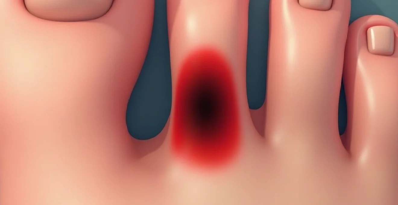
Subungual haematoma: Trauma-Induced nail bed bleeding
Subungual haematomas represent the most frequent cause of black spots under toenails, accounting for approximately 85% of all cases presenting to podiatry clinics. This condition occurs when blood accumulates between the nail plate and underlying nail bed following trauma. The trapped blood creates a characteristic dark purple to black discolouration that can vary in size from small spots to complete nail coverage.
The pathophysiology involves disruption of capillary networks within the nail bed, leading to extravasation of blood into the subungual space. Unlike surface bruising that resolves relatively quickly , subungual haematomas remain trapped beneath the rigid nail plate, creating persistent discolouration. The confined space can generate significant pressure, particularly in acute cases, leading to throbbing pain that intensifies with dependency or physical activity.
Acute injury mechanisms and blood accumulation patterns
Acute subungual haematomas typically develop within 24-48 hours of injury, presenting with immediate pain and progressive darkening. The blood accumulation pattern often reflects the injury mechanism, with crush injuries creating more diffuse bleeding compared to penetrating trauma that produces localised spots. Understanding these patterns helps differentiate fresh haematomas from chronic conditions .
The volume of blood accumulation directly correlates with symptom severity and treatment requirements. Small haematomas involving less than 25% of the nail surface usually resolve without intervention, while larger collections may require drainage to relieve pressure and prevent nail loss. Progressive enlargement of the dark area within the first few days suggests ongoing bleeding that may benefit from medical evaluation.
Door crush injuries and heavy object impact assessment
Door crush injuries represent a particularly common mechanism for severe subungual haematomas, often occurring when fingers or toes become trapped in closing doors. The sudden compressive force can cause extensive nail bed damage, resulting in large haematomas that may occupy the entire nail surface. These injuries frequently produce the most painful presentations due to rapid pressure accumulation.
Heavy object impacts, such as dropped tools or furniture, create different injury patterns characterised by focal areas of intense bleeding. The force distribution determines whether the haematoma remains localised or spreads across the nail bed. Assessment of the injury mechanism helps predict healing time and potential complications , including nail loss or permanent deformity.
Sports-related toenail trauma in running and football
Athletic activities contribute significantly to subungual haematoma development, with running and football presenting the highest incidence rates. Repetitive microtrauma from ill-fitting footwear, combined with the repetitive stress of athletic movement, creates conditions conducive to nail bed bleeding. Studies indicate that up to 30% of marathon runners experience some degree of toenail discolouration during training or competition.
Football players face additional risks from direct kicks, studs contact, and rapid direction changes that stress the nail-shoe interface. The combination of moisture, pressure, and repetitive trauma creates an environment where small amounts of bleeding can accumulate over time, producing gradual darkening rather than acute onset symptoms. Recognition of sports-related patterns helps athletes modify training or equipment to prevent recurrence .
Subungual pressure symptoms and pain management
Pain intensity in subungual haematomas varies considerably based on the volume of blood accumulation and rate of development. Acute haematomas often produce severe, throbbing pain that worsens with activity or dependent positioning. The rigid nail plate prevents swelling accommodation, creating pressure that can reach levels sufficient to compromise local blood flow.
Clinical studies demonstrate that haematomas involving more than 50% of the nail surface frequently require intervention to prevent complications and provide symptom relief.
Conservative pain management includes elevation, ice application, and oral analgesics for mild to moderate discomfort. However, severe pain or progressive symptoms may indicate the need for nail trephination, a procedure where small holes are created in the nail to drain accumulated blood and relieve pressure. This intervention provides immediate pain relief and can preserve nail integrity when performed appropriately.
Malignant melanoma under toenails: acral lentiginous melanoma
Subungual melanoma represents one of the most serious causes of nail discolouration, though it accounts for less than 2% of all nail discolouration cases. This aggressive form of skin cancer typically presents as acral lentiginous melanoma, a subtype that disproportionately affects individuals with darker skin tones. Unlike other melanoma types, subungual melanoma shows no correlation with sun exposure, making it particularly insidious as it develops in areas rarely examined during routine skin checks.
The incidence of subungual melanoma varies significantly across ethnic groups, with rates of 15-20% among African Americans compared to 2-3% in Caucasian populations. This cancer typically affects the thumb or great toe, areas subject to repeated minor trauma that may contribute to malignant transformation. Early detection remains crucial for optimal outcomes , as advanced cases carry poor prognoses due to late presentation and rapid metastatic potential.
Hutchinson’s sign and pigmented nail streaks
Hutchinson’s sign represents one of the most reliable indicators of subungual melanoma, characterised by extension of pigmentation from the nail onto the surrounding skin (periungual tissue). This finding suggests that the pigmented lesion has grown beyond the confines of the nail unit, indicating a more advanced stage of disease. The presence of Hutchinson’s sign warrants immediate dermatological evaluation and typically necessitates urgent biopsy.
Pigmented nail streaks associated with melanoma typically display irregular borders, varying width, and progressive darkening over time. Unlike benign pigmentation that remains stable, malignant lesions demonstrate dynamic changes in size, shape, and colour intensity. The longitudinal melanonychia may start as a narrow band but progressively widens, often developing areas of different pigmentation intensity within the same streak.
ABCDE criteria for subungual melanoma detection
The traditional ABCDE criteria for melanoma detection require modification when applied to nail lesions, as conventional asymmetry and border irregularity assessments prove challenging in the nail environment. Adapted criteria focus on Age (typically over 50), Band characteristics (brown-black colour, width >3mm, irregular borders), Change in the nail appearance, Digit involvement (thumb or great toe most commonly), and Extension onto periungual skin.
Colour variation within nail streaks provides particularly important diagnostic information, with melanomas often displaying multiple shades of brown, black, or even amelanotic (non-pigmented) areas. Progressive widening of pigmented bands , especially at the proximal nail fold, strongly suggests malignant transformation. The rate of change serves as a critical factor, with melanomas typically demonstrating noticeable progression over weeks to months rather than remaining stable over years.
Dermoscopic features of nail unit melanoma
Dermoscopy enhances diagnostic accuracy for nail lesions, revealing microscopic features invisible to naked eye examination. Malignant lesions often display irregular pigment patterns, including micro-Hutchinson’s sign (periungual extension visible only under magnification) and colour variegation within the nail streak. Advanced dermoscopic techniques can identify subtle features that distinguish melanoma from benign conditions.
Specific dermoscopic patterns associated with subungual melanoma include irregular parallel ridges, background discolouration, and disruption of normal nail plate architecture. These features contrast with the regular parallel ridge pattern typically seen in benign longitudinal melanonychia. Dermoscopic examination significantly improves diagnostic confidence and helps guide decisions regarding biopsy necessity.
Differential diagnosis between melanoma and benign pigmentation
Distinguishing subungual melanoma from benign pigmentation challenges even experienced clinicians, as both conditions can present with similar appearance initially. Benign longitudinal melanonychia typically remains stable over time, displays regular borders, and rarely exceeds 3mm in width. Cultural and genetic factors influence the prevalence of benign pigmentation, with up to 77% of African Americans demonstrating some degree of nail pigmentation.
Recent advances in nail biopsy techniques have improved diagnostic accuracy while minimising cosmetic impact, enabling definitive diagnosis when clinical features remain ambiguous.
Trauma history can complicate differential diagnosis, as injury may trigger pigmentation in previously normal nails or alter the appearance of existing lesions. However, post-traumatic pigmentation typically shows gradual fading over months, while melanomas demonstrate progressive changes. Age represents another discriminating factor, with melanoma predominantly affecting individuals over 40, while benign pigmentation commonly begins in childhood or adolescence.
Fungal infections causing dark nail discolouration
Onychomycosis, or fungal nail infection, ranks as the second most common cause of nail discolouration, affecting approximately 10-15% of the population. While yellow and white discolouration represents the classic presentation, certain fungal species and secondary bacterial infections can produce dark brown to black pigmentation. These infections typically develop gradually over months to years, accompanied by progressive nail thickening, brittleness, and structural changes.
The pathophysiology involves fungal invasion of the nail plate and underlying structures, leading to keratin degradation and inflammatory responses. Specific fungal species demonstrate preferential pigment production , creating characteristic colour patterns that aid in species identification. Environmental factors, including humidity, warmth, and compromised nail integrity, predispose individuals to infection and influence the clinical presentation.
Onychomycosis with pseudomonas aeruginosa secondary infection
Secondary bacterial infections complicate approximately 20% of chronic fungal nail infections, with Pseudomonas aeruginosa representing the most common bacterial pathogen. This gram-negative bacterium produces distinctive blue-green to black pigmentation through pyocyanin and pyoverdine production. The combination of fungal structural damage and bacterial colonisation creates ideal conditions for rapid pigment development.
Pseudomonas superinfection typically occurs in chronically damaged nails with compromised barrier function. The bacterial infection often produces a characteristic odour, described as sweet or grape-like, which helps differentiate it from purely fungal infections. Prompt recognition of bacterial superinfection requires systemic antibiotic therapy in addition to antifungal treatment to prevent serious complications.
Aspergillus niger and black nail syndrome
Aspergillus niger infections, though relatively rare, produce distinctive black discolouration that can mimic subungual haematomas or melanoma. This saprophytic fungus thrives in humid environments and typically affects immunocompromised individuals or those with extensive nail damage. The infection produces characteristic black spores that create punctate or diffuse darkening patterns.
Black nail syndrome associated with Aspergillus niger often affects multiple nails simultaneously, helping distinguish it from traumatic haematomas that typically involve single digits. The infection demonstrates progressive spread if untreated, with darkening advancing from the distal nail edge proximally. Antifungal susceptibility testing guides appropriate therapy , as Aspergillus species show variable sensitivity to standard antifungal agents.
Candida parapsilosis dark pigment production
Candida parapsilosis represents an emerging cause of dark nail discolouration, particularly in healthcare workers and individuals with frequent water exposure. This yeast species demonstrates unusual pigment production capabilities under specific environmental conditions, creating brown to black discolouration that can confuse clinical diagnosis. The infection often affects the nail fold initially before spreading to involve the entire nail unit.
Clinical presentation typically includes painful paronychia (nail fold inflammation) accompanied by progressive nail discolouration and structural changes. The combination of yeast infection and pigment production creates a unique clinical picture that requires specific antifungal therapy. Recognition of this pattern helps avoid misdiagnosis and ensures appropriate treatment selection.
KOH testing and fungal culture identification methods
Potassium hydroxide (KOH) preparation remains the primary diagnostic tool for suspected fungal nail infections, providing rapid results that guide initial treatment decisions. The test demonstrates fungal elements including hyphae and spores, confirming the presence of infection. However, KOH testing cannot differentiate between fungal species or identify concurrent bacterial infections that may contribute to discolouration.
Fungal culture provides definitive species identification and antifungal susceptibility information, though results require 2-6 weeks for completion. Culture-directed therapy improves treatment success rates and helps identify resistant organisms that may require alternative therapeutic approaches. Modern molecular diagnostic techniques, including PCR-based methods, offer rapid species identification with improved sensitivity compared to traditional culture methods.
Bacterial infections and pseudomonas nail colonisation
Bacterial nail infections, particularly those involving Pseudomonas aeruginosa, create distinctive green-black discolouration through bacterial pigment production. These infections typically develop in compromised nail environments, including chronic moisture exposure, nail damage, or concurrent fungal infections. The bacterial colonisation produces characteristic pigments including pyocyanin (blue-green) and pyoverdine (yellow-green) that combine to create dark discolouration.
Pseudomonas nail infections demonstrate rapid progression compared to fungal infections, with visible colour changes developing over days to weeks rather than months. The bacteria thrive in moist environments and can quickly colonise damaged nail tissue, producing biofilms that enhance antibiotic resistance. Early recognition and treatment prevent serious complications including cellulitis, abscess formation, and potential systemic infection in immunocompromised individuals.
Risk factors for bacterial nail colonisation include occupational water exposure, nail trauma, diabetes mellitus, and immunosuppression. Healthcare workers, food service employees, and individuals with hobbies involving water exposure show increased susceptibility. The infection often begins in areas of nail damage or separation, spreading to involve larger nail areas if untreated. Clinical presentation includes rapid onset discolouration, distinctive odour, and potential pain or tenderness around the affected nail.
Treatment requires appropriate antibiotic selection based on culture and sensitivity results, as Pseudomonas demonstrates inherent resistance to many common antibiotics. Topical antiseptics and systemic antibiotics may both be necessary for effective treatment. Prevention focuses on maintaining nail hygiene, avoiding prolonged moisture exposure, and promptly treating nail injuries that could provide bacterial entry points.
Drug-induced melanonychia and medication side effects
Medication-induced nail pigmentation represents an underrecognised cause of dark nail discolouration, affecting up to 40% of patients receiving certain systemic therapies. These pigmentary changes result from drug accumulation in nail tissues, stimulation of melanin production, or direct chemical reactions within the nail unit. Recognition of drug-induced changes prevents unnecessary anxiety and inappropriate interventions while the underlying condition continues treatment.
The mechanism of drug-induced melanonychia varies by medication class and individual patient factors. Some drugs stimulate melanocyte activity within the nail matrix, producing longitudinal pigmented bands similar to those seen in ethnic pigmentation. Others create chemical reactions that produce dark compounds within the nail structure, resulting in diffuse discolouration. The timing of onset typically correlates with drug initiation, though some medications require months of therapy before visible changes appear.
Chemotherapy-related nail pigmentation changes
Chemotherapy agents frequently cause nail pigmentation changes, with incidence rates varying from 12% to 74% depending on the specific drug regimen. Common agents including doxorubicin, bleomycin, and cyclophosphamide produce characteristic patterns ranging from diffuse darkening to distinct horizontal bands corresponding to treatment cycles. These changes typically resolve gradually following treatment completion , though some pigmentation may persist permanently.
The characteristic banding pattern often coincides with treatment schedules, creating a timeline of therapy visible in nail growth. Patients receiving intensive chemotherapy protocols may develop multiple bands representing different treatment cycles. These pigmentary changes can affect fingernails and toenails simultaneously, though toenails often show more pronounced discolouration due to slower growth rates that concentrate drug exposure.
Management focuses on patient education and reassurance, as chemotherapy-induced nail changes rarely require intervention. Protective nail care during treatment can minimise severity, including gentle handling, moisturising, and avoiding trauma. Some oncologists recommend nail cooling during chemotherapy infusion to reduce drug uptake and subsequent pigmentation, though evidence remains limited.
Antimalarial drug hyperpigmentation effects
Antimalarial medications, particularly hydroxychloroquine and chloroquine, produce distinctive nail pigmentation patterns in approximately 25% of long-term users. The discolouration typically appears as blue-grey to black bands or diffuse darkening that develops gradually over months of therapy. These changes reflect drug accumulation in melanin-rich tissues, including the nail matrix where melanocytes produce pigment.
The pigmentation pattern often correlates with cumulative drug exposure, with higher doses and longer treatment durations producing more pronounced changes. Patients with autoimmune conditions requiring long-term antimalarial therapy show the highest incidence of nail discolouration. The changes typically stabilise once steady-state drug levels are achieved, though complete resolution may require years following discontinuation.
Clinical presentation includes longitudinal streaking or diffuse nail darkening that may affect multiple digits simultaneously. Unlike melanoma, drug-induced changes typically show bilateral symmetry and stable progression patterns. Documentation of medication history prevents misdiagnosis and unnecessary invasive procedures. Healthcare providers should counsel patients about potential cosmetic changes before initiating long-term antimalarial therapy.
Zidovudine and HIV medication nail discolouration
Zidovudine (AZT) and other nucleoside reverse transcriptase inhibitors commonly cause nail pigmentation changes in HIV-positive patients, with reported incidence rates reaching 40% in some studies. The discolouration typically manifests as longitudinal blue-black bands or diffuse darkening that develops within weeks to months of treatment initiation. These changes occur more frequently in patients with darker skin tones, suggesting interaction with existing melanin production pathways.
The mechanism involves drug interference with DNA synthesis in rapidly dividing nail matrix cells, leading to abnormal melanin deposition and cellular damage. Multiple nails are typically affected simultaneously, creating a characteristic pattern that helps distinguish drug-induced changes from other causes of nail discolouration. The pigmentation may intensify with continued therapy but rarely causes functional impairment.
Studies indicate that nail pigmentation changes may serve as visible markers of drug compliance and absorption in HIV patients, providing clinicians with additional monitoring parameters during antiretroviral therapy.
Management involves patient education about expected changes and monitoring for signs of drug toxicity. While cosmetically concerning, these pigmentary changes rarely necessitate treatment discontinuation unless accompanied by other serious adverse effects. The discolouration typically stabilises with continued therapy and may gradually fade following drug discontinuation, though complete resolution can require 12-18 months due to slow nail growth rates.