Distinguishing between a benign blood blister and potentially malignant skin cancer represents one of the most challenging diagnostic dilemmas in dermatological practice. The visual similarity between certain haemorrhagic lesions and pigmented malignancies can create significant anxiety for patients and occasionally perplex even experienced clinicians. Understanding the fundamental differences between these conditions becomes crucial when you consider that early detection of skin cancer dramatically improves treatment outcomes, whilst unnecessary concern over harmless blood blisters can cause undue stress. This diagnostic complexity is particularly pronounced with nodular melanoma, which can closely mimic the appearance of traumatic haemorrhagic vesicles. The ability to recognise key distinguishing features enables more informed decision-making about when to seek professional medical evaluation.
Understanding blood blisters: pathophysiology and clinical presentation
Haemorrhagic vesicle formation mechanisms in dermal tissue
Blood blisters develop through a well-defined pathophysiological process involving mechanical trauma to cutaneous blood vessels without full-thickness skin disruption. When sufficient force is applied to the dermis, capillaries and small venules rupture beneath an intact epidermis, allowing extravasated blood to accumulate within the dermal-epidermal junction. This process differs fundamentally from standard friction blisters, where serous fluid rather than blood fills the cavity. The integrity of the overlying epidermis prevents immediate blood loss whilst creating the characteristic raised, blood-filled lesion that defines this condition.
The formation mechanism typically involves shearing forces that exceed the tensile strength of dermal blood vessels. Pinching injuries represent the most common causative mechanism, where opposing forces compress tissue sufficiently to rupture vascular structures. The resulting haematoma becomes encapsulated by the natural tissue planes, creating the distinctive bubble-like appearance. Understanding this mechanism helps explain why blood blisters predominantly occur in areas subject to mechanical stress or trauma.
Characteristic morphological features of subepidermal haematomas
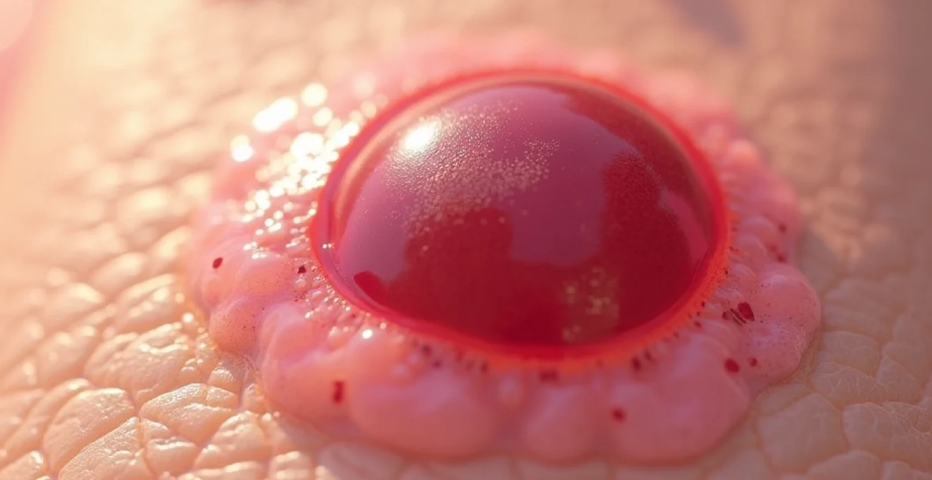
The morphological characteristics of blood blisters follow predictable patterns that aid in clinical recognition. These lesions typically present as well-demarcated, raised vesicles containing dark red to purple fluid. The overlying epidermis remains intact but appears stretched and translucent, allowing visualisation of the contained haematoma. Size varies considerably depending on the extent of vascular damage and local tissue characteristics, ranging from a few millimetres to several centimetres in diameter.
The colour evolution of blood blisters provides important diagnostic information. Fresh lesions display bright red colouration due to oxygenated haemoglobin, gradually transitioning through darker red and purple phases as deoxygenation occurs. Over time, haemoglobin breakdown products create characteristic colour changes, progressing through blue-green phases before eventual resolution. This predictable colour evolution differs markedly from the stable pigmentation patterns observed in melanocytic malignancies.
Common anatomical locations for traumatic blood blister development
Blood blisters demonstrate distinct predilection sites that correlate directly with mechanical stress patterns and trauma susceptibility. The digits represent the most frequent location, particularly the fingertips and areas adjacent to nail beds where pinching injuries commonly occur. Palmar and plantar surfaces also show increased susceptibility due to repetitive friction and pressure during daily activities. These anatomical preferences reflect the mechanical nature of blood blister formation.
Within the oral cavity, blood blisters frequently develop on the tongue, buccal mucosa, and lip margins following accidental bite injuries. The thin, vascular nature of oral mucosa makes these tissues particularly susceptible to haemorrhagic vesicle formation. Recognition of these common distribution patterns helps distinguish traumatic blood blisters from malignant lesions, which may occur in sun-exposed areas or follow different anatomical preferences depending on the specific cancer type.
Duration and natural history of haemorrhagic blisters
The natural history of blood blisters follows a predictable timeline that provides valuable diagnostic information. Most uncomplicated lesions resolve spontaneously within 7-14 days through natural reabsorption of extravasated blood and tissue remodelling processes. The overlying epidermis may remain intact throughout this period, or may eventually desquamate as healing progresses. Resolution occurs without scarring in the vast majority of cases, leaving normal skin architecture once healing is complete.
Factors influencing healing duration include lesion size, anatomical location, and individual patient characteristics such as age and vascular health. Larger lesions naturally require extended healing periods, whilst locations subject to continued mechanical stress may experience delayed resolution. Understanding these temporal patterns becomes crucial when differentiating blood blisters from malignant lesions, which typically demonstrate progressive growth rather than spontaneous resolution.
Melanoma and Non-Melanoma skin cancer: diagnostic characteristics
ABCDE criteria assessment for malignant melanoma detection
The ABCDE mnemonic represents the gold standard for melanoma recognition, providing a systematic approach to evaluating suspicious pigmented lesions. Asymmetry assessment involves examining whether one half of a lesion mirrors the other when divided by an imaginary central line. Malignant melanomas frequently demonstrate marked asymmetry, contrasting with the typically symmetrical appearance of benign naevi. This asymmetry often represents the first detectable sign of malignant transformation in melanocytic lesions.
Border irregularity constitutes another critical diagnostic feature, with malignant lesions typically displaying uneven, scalloped, or notched margins rather than the smooth, regular borders characteristic of benign moles. Colour variation within a single lesion raises significant concern, particularly when multiple hues including black, brown, red, white, or blue appear within the same growth. The diameter criterion traditionally focused on lesions exceeding 6mm, though contemporary understanding recognises that smaller melanomas can be equally dangerous.
Early-stage melanomas may not conform to all traditional ABCDE criteria, making clinical vigilance and professional evaluation essential for any changing or concerning lesions.
Basal cell carcinoma: nodular and superficial variants recognition
Basal cell carcinoma presents in multiple morphological variants that can occasionally mimic haemorrhagic lesions, particularly in the nodular form. The classic nodular basal cell carcinoma appears as a pearly, translucent papule with visible telangiectasias and a tendency toward central ulceration. The “rolled” or raised border represents a pathognomonic feature that helps distinguish this malignancy from benign lesions. These tumours grow slowly and rarely metastasise, though local destruction can be significant if treatment is delayed.
Superficial basal cell carcinomas present as erythematous, scaling patches that may be confused with benign inflammatory conditions. These lesions typically occur on the trunk and demonstrate gradual enlargement over months to years. The presence of a subtle raised border and resistance to topical treatments often provides diagnostic clues. Recognition of these variants becomes important when evaluating any persistent lesion that fails to heal or continues growing despite conservative management.
Squamous cell carcinoma: keratotic and ulcerative presentations
Squamous cell carcinoma manifests in various forms that can sometimes be confused with traumatic lesions, particularly when ulceration is present. The keratotic variant presents as a hyperkeratotic, rough-surfaced papule or plaque with a tendency toward scale formation. These lesions often develop in sun-damaged skin and may arise from pre-existing actinic keratoses. The firm, indurated quality typically distinguishes these malignancies from softer, more compressible blood blisters.
Ulcerative squamous cell carcinomas present as non-healing wounds with raised, everted edges and granulation tissue in the base. These lesions may bleed easily and fail to respond to conventional wound care measures. The persistent nature of these ulcers, combined with their progressive enlargement, contrasts markedly with the self-limiting course of traumatic blood blisters. Historical context and patient recall of initial lesion appearance often provide valuable diagnostic information.
Amelanotic melanoma: identifying Pigment-Free malignant lesions
Amelanotic melanomas represent particularly challenging diagnostic entities due to their lack of characteristic pigmentation, making them easily confused with benign lesions including blood blisters. These aggressive malignancies may appear as pink, red, or flesh-coloured nodules that grow rapidly and may bleed or ulcerate. The absence of melanin removes the typical visual cues that alert clinicians to melanocytic malignancy, often leading to delayed diagnosis and worse outcomes.
Key distinguishing features of amelanotic melanoma include rapid growth, firm consistency, and tendency toward bleeding or ulceration without obvious trauma. The ABCDE criteria prove less reliable for these lesions, making clinical suspicion and low threshold for biopsy essential. Any rapidly growing, non-pigmented lesion that bleeds easily or fails to heal should prompt immediate dermatological evaluation, regardless of its resemblance to benign conditions.
Kaposi sarcoma and angiosarcoma: vascular malignancy appearances
Vascular malignancies can closely mimic haemorrhagic blisters due to their inherent vascularity and tendency toward bleeding. Kaposi sarcoma typically presents as purple to red-brown patches, plaques, or nodules that may resemble organised haematomas. The multifocal nature of Kaposi sarcoma often provides diagnostic clues, as multiple lesions typically appear simultaneously in susceptible patients, contrasting with the usually solitary nature of traumatic blood blisters.
Angiosarcoma represents an aggressive vascular malignancy that may present as rapidly enlarging, haemorrhagic nodules or plaques. These lesions often occur in chronically lymphoedematous tissue or previously irradiated areas. The rapid progression and tendency toward local tissue destruction distinguish angiosarcomas from benign haemorrhagic lesions. Clinical context, including medical history and anatomical location, often provides crucial diagnostic information for these rare but aggressive malignancies.
Differential diagnosis using dermoscopic evaluation techniques
Dermoscopy patterns in benign haemorrhagic lesions
Dermoscopic examination of blood blisters reveals characteristic features that distinguish them from malignant lesions. The most prominent finding involves homogeneous red-to-purple colouration without internal structure, reflecting the uniform distribution of extravasated blood within the dermal space. Absence of pigment network , streaks, or globules helps exclude melanocytic processes. The sharp demarcation between the haemorrhagic area and surrounding normal skin provides another important diagnostic clue.
Under polarised dermoscopy, blood blisters typically demonstrate a uniform appearance without the complex vascular patterns seen in malignant lesions. The contained nature of the haematoma prevents the irregular vessel proliferation characteristic of neoplastic processes. Sequential dermoscopic examination can document the expected evolution of colour changes as haemoglobin breakdown occurs, providing additional diagnostic confidence when clinical uncertainty exists.
Vascular structures analysis in malignant skin tumours
Malignant lesions demonstrate characteristic vascular patterns under dermoscopic examination that differ significantly from the uniform appearance of blood blisters. Atypical vascular structures including dotted, linear irregular, and hairpin vessels often appear within malignant lesions, reflecting the abnormal angiogenesis associated with neoplastic growth. These patterns contrast markedly with the homogeneous blood pool seen in traumatic haemorrhagic lesions.
Melanomas may display multiple vascular morphologies within a single lesion, reflecting the heterogeneous nature of malignant tissue. Basal cell carcinomas typically show fine telangiectatic vessels arranged in tree-like patterns, whilst squamous cell carcinomas often demonstrate glomerular or hairpin vessel configurations. Recognition of these specific vascular signatures aids in distinguishing malignant processes from benign haemorrhagic lesions during clinical evaluation.
Pigment network assessment and melanocytic lesion identification
The presence of pigment network represents a key dermoscopic feature that helps identify melanocytic lesions and distinguish them from haemorrhagic blisters. Normal melanocytic naevi demonstrate regular, honeycomb-like pigment networks with uniform hole size and line thickness. Atypical networks with irregular holes, variable line thickness, or abrupt termination raise concern for malignant transformation. Blood blisters lack any pigment network structure, showing only uniform colouration from contained blood.
Advanced dermoscopic features such as blue-white veil, regression structures, and peripheral streaks indicate melanocytic proliferation and potential malignancy. These sophisticated patterns never appear in simple haemorrhagic lesions, providing clear diagnostic differentiation. The ability to recognise and interpret these pigmentary patterns represents a crucial skill in distinguishing between benign haemorrhagic lesions and potentially dangerous melanocytic malignancies.
Border irregularity and asymmetry evaluation methods
Dermoscopic assessment of border characteristics provides valuable information for distinguishing malignant lesions from blood blisters. Malignant lesions typically demonstrate irregular borders with varying degrees of indentation, protrusion, and scalloping. The border assessment requires systematic evaluation of the entire lesion perimeter, noting any areas of irregular outline or abrupt colour transitions. Blood blisters generally maintain smooth, regular borders that reflect the mechanical forces involved in their formation.
Asymmetry evaluation involves comparing corresponding areas of a lesion to identify discordant features. Mathematical approaches using grid overlays can quantify asymmetry objectively, though clinical pattern recognition often suffices for practical diagnosis. The uniform nature of blood blisters typically results in symmetrical lesions, contrasting with the inherent asymmetry common in malignant growths. This systematic approach to border and asymmetry assessment enhances diagnostic accuracy when evaluating suspicious lesions.
Clinical red flags requiring immediate dermatological referral
Several clinical features warrant urgent dermatological evaluation when encountered in any skin lesion, regardless of initial clinical impression. Rapid growth represents perhaps the most concerning feature, particularly when a lesion doubles in size within weeks to months. This growth pattern strongly suggests malignant potential and differs markedly from the static or slowly resolving nature of typical blood blisters. Any lesion demonstrating this aggressive growth pattern requires immediate professional assessment.
Spontaneous bleeding without trauma indicates tissue fragility and possible malignant transformation. Whilst blood blisters may rupture and bleed when subjected to pressure or friction, spontaneous bleeding suggests underlying pathological processes. Ulceration or non-healing wounds that persist beyond expected healing timeframes raise significant concern for malignant disease. The combination of bleeding and ulceration in a rapidly growing lesion represents a dermatological emergency requiring urgent evaluation.
Any lesion that bleeds spontaneously, grows rapidly, or fails to heal within expected timeframes requires prompt professional evaluation, regardless of its initial appearance or suspected benign nature.
Changes in sensation, including new onset of itching, pain, or numbness, may indicate malignant infiltration of neural structures. Blood blisters typically cause discomfort related to mechanical pressure but rarely produce the neurological symptoms associated with invasive malignancies. Satellite lesions or regional lymph node enlargement suggest possible metastatic disease and mandate immediate oncological assessment. These systemic signs differentiate aggressive malignancies from localised benign processes.
The development of multiple similar lesions in different anatomical regions raises concern for systemic disease processes, including disseminated malignancies or paraneoplastic phenomena. This pattern contrasts with the typically solitary nature of traumatic blood blisters. Additionally, lesions occurring in patients with immunocompromise, previous radiation exposure, or significant family history of skin cancer warrant lower thresholds for professional evaluation due to increased malignant potential.
Professional diagnostic methods: biopsy and histopathological analysis
When clinical examination cannot definitively distinguish between blood blisters and malignant lesions, tissue sampling becomes necessary for accurate diagnosis. Punch biopsy represents the most commonly employed technique for small, suspicious lesions, providing adequate tissue for histopathological analysis whilst minimising procedural complexity. The technique involves using a circular cutting instrument to obtain a full-thickness skin sample including epidermis, dermis, and superficial subcutaneous tissue.
Excisional biopsy may be preferred for lesions
where suspicion of malignancy exists, as this approach provides complete lesion removal for comprehensive histological assessment. This method proves particularly valuable when dealing with pigmented lesions where complete architectural evaluation is essential for accurate diagnosis. Incisional biopsy may be appropriate for large lesions where complete excision would result in significant cosmetic or functional impairment.
The histopathological analysis of suspected blood blisters reveals characteristic features that confirm the diagnosis and exclude malignant processes. Microscopic examination typically demonstrates extravasated red blood cells within the dermal-epidermal space, accompanied by variable degrees of acute inflammation. The overlying epidermis remains intact, though it may show reactive changes including spongiosis or mild hyperplasia. Absence of atypical cellular morphology or abnormal proliferation patterns helps definitively exclude malignant processes.
Immunohistochemical staining may be employed when conventional histological features prove insufficient for definitive diagnosis. Markers such as S-100 and Melan-A help identify melanocytic lesions, whilst cytokeratin stains assist in characterising epithelial malignancies. These sophisticated diagnostic tools become particularly valuable when evaluating amelanotic lesions or poorly differentiated malignancies that may lack characteristic morphological features on routine staining.
Management protocols for blood blisters versus suspected malignancies
The management approach for confirmed blood blisters emphasises conservative care focused on preventing secondary complications whilst allowing natural healing processes to occur. Protection from further trauma represents the cornerstone of blood blister management, typically achieved through appropriate padding, bandaging, or activity modification. The intact overlying epidermis should be preserved whenever possible, as it provides an excellent biological dressing that promotes healing and prevents infection.
Drainage of blood blisters remains controversial, with most dermatologists recommending against routine aspiration due to infection risk and minimal symptomatic benefit. When drainage becomes necessary due to significant discomfort or functional impairment, sterile technique must be employed with careful attention to maintaining the overlying skin flap. Topical antibiotic prophylaxis may be considered following drainage procedures, particularly in immunocompromised patients or those with diabetes mellitus.
Pain management for blood blisters typically involves simple analgesics such as paracetamol or non-steroidal anti-inflammatory drugs, depending on patient tolerance and contraindications. Cold compresses may provide symptomatic relief during the acute phase, whilst elevation of affected extremities can help reduce swelling and associated discomfort. Patients should be educated about expected healing timeframes and signs of secondary infection that would warrant medical re-evaluation.
Suspected malignant lesions require urgent referral to appropriate specialists, with timeframes dependent on clinical suspicion level and available resources. Two-week wait pathways are typically utilised for suspected skin cancers in many healthcare systems, ensuring rapid access to specialist evaluation and definitive management. The referral process should include comprehensive clinical documentation, including lesion photography when possible, to facilitate appropriate triaging and treatment planning.
Following malignancy diagnosis, multidisciplinary team involvement becomes essential for optimal treatment planning. Surgical excision remains the primary treatment modality for most skin cancers, with margin requirements varying according to histological subtype and staging. Mohs micrographic surgery may be preferred for certain high-risk lesions or those in cosmetically sensitive areas where tissue preservation is crucial. Adjuvant therapies including radiotherapy, immunotherapy, or targeted molecular treatments may be indicated for advanced cases.
Long-term surveillance protocols differ significantly between blood blister patients and those with confirmed malignancies. Patients with simple blood blisters require minimal follow-up beyond ensuring complete resolution and providing education about prevention strategies. Cancer patients enter structured surveillance programmes with regular clinical examinations, imaging studies when indicated, and patient education regarding self-examination techniques for detecting potential recurrence or new primary lesions.
Prevention strategies for recurrent blood blisters focus on identifying and modifying causative factors through lifestyle adjustments, protective equipment use, and environmental modifications. Patients should be counselled regarding appropriate footwear selection, workplace safety measures, and recognition of early warning signs that might prevent lesion development. For malignant conditions, prevention emphasises sun protection measures, regular skin examinations, and genetic counselling when indicated for patients with strong family histories of skin cancer.