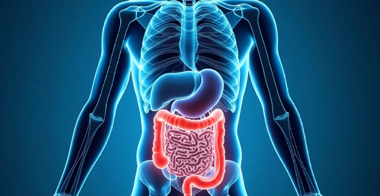
Diarrhoea following computed tomography (CT) scans with contrast media represents one of the most frequently reported gastrointestinal side effects in diagnostic imaging. This physiological response affects approximately 15-25% of patients undergoing contrast-enhanced CT procedures, ranging from mild, transient loose stools to more significant digestive disturbances requiring clinical intervention. Understanding the mechanisms behind this common adverse reaction becomes essential for both healthcare providers and patients preparing for diagnostic imaging procedures.
The occurrence of post-contrast diarrhoea stems from complex interactions between contrast agents and the gastrointestinal tract, involving osmotic effects, alterations in intestinal motility, and potential disruption of the gut microbiome. While generally considered a normal physiological response, recognising when symptoms warrant medical attention remains crucial for patient safety and optimal care outcomes.
Understanding contrast media composition and gastrointestinal impact
Modern contrast agents used in CT imaging comprise sophisticated chemical compounds designed to enhance tissue differentiation whilst minimising adverse effects. The three primary categories of contrast media—iodinated compounds, barium-based preparations, and gadolinium agents—each interact differently with the gastrointestinal system, producing varying degrees of digestive side effects.
The molecular structure and osmolality of these agents significantly influence their propensity to cause gastrointestinal disturbances. Osmolality , measured in milliosmoles per kilogram of water, determines how contrast media affects fluid balance within the intestinal tract. Higher osmolality solutions tend to draw more water into the bowel lumen, potentially triggering loose stools or diarrhoea through osmotic mechanisms.
Iodinated contrast agents: iohexol and iopamidol properties
Iohexol and iopamidol represent the most commonly employed non-ionic, low-osmolar contrast media in contemporary clinical practice. These agents possess osmolality values ranging from 290 to 320 mOsm/kg H₂O, significantly lower than earlier ionic preparations that often exceeded 1500 mOsm/kg H₂O. Despite this improvement, approximately 12-18% of patients still experience gastrointestinal symptoms following intravenous administration.
The gastrointestinal impact of these agents occurs through multiple pathways. When administered intravenously, small quantities of iodinated contrast inevitably reach the intestinal tract through normal physiological processes. The presence of these compounds within the bowel lumen can alter water absorption patterns and stimulate increased peristaltic activity, contributing to loose stool formation.
Osmolality effects of Low-Osmolar contrast media on intestinal function
Low-osmolar contrast media, whilst representing a significant advancement in patient tolerance, still exert measurable effects on intestinal physiology. The osmotic gradient created by these agents within the bowel lumen can result in osmotic diarrhoea , characterised by increased stool volume and frequency without significant inflammatory components.
Clinical studies demonstrate that even minimal osmolality differences can influence gastrointestinal outcomes. Patients receiving iso-osmolar agents (approximately 290 mOsm/kg H₂O) report digestive disturbances in 8-12% of cases, compared to 15-20% for slightly hyper-osmolar preparations. This dose-response relationship underscores the importance of osmolality in determining gastrointestinal tolerability.
Barium sulphate versus iodinated contrast: digestive system interactions
Barium sulphate preparations, primarily used for gastrointestinal tract imaging, interact more directly with the digestive system due to their oral or rectal administration routes. These agents create a coating effect within the bowel, potentially altering normal digestive processes and bacterial interactions. The incidence of post-procedure diarrhoea following barium studies ranges from 20-35%, notably higher than intravenous iodinated contrast administration.
The persistence of barium within the gastrointestinal tract for 24-72 hours post-procedure contributes to prolonged digestive effects. Unlike water-soluble iodinated agents that rapidly clear from the system, barium’s extended residence time can result in sustained alterations to bowel function, including both diarrhoea and constipation patterns.
Gadolinium-based contrast agents and rare gastrointestinal reactions
Gadolinium-based contrast agents, primarily utilised in magnetic resonance imaging, occasionally cause gastrointestinal disturbances when small amounts are eliminated through biliary pathways. The incidence of diarrhoea following gadolinium administration remains relatively low at 3-7%, reflecting the agent’s predominantly renal elimination pathway and minimal direct gastrointestinal exposure.
When gastrointestinal symptoms do occur with gadolinium agents, they typically manifest as mild nausea or loose stools rather than significant diarrhoea. The chelated structure of these compounds reduces their interaction with intestinal receptors, contributing to their favourable gastrointestinal tolerability profile.
Physiological mechanisms behind Post-Contrast diarrhoea
The development of diarrhoea following contrast media administration involves multiple, interconnected physiological pathways that extend beyond simple osmotic effects. These mechanisms operate simultaneously and synergistically, creating a complex cascade of events that ultimately result in altered bowel function and increased stool frequency.
Osmotic diarrhoea pathophysiology following contrast administration
Osmotic diarrhoea represents the primary mechanism underlying post-contrast gastrointestinal symptoms. When contrast agents enter the intestinal lumen, either through direct oral administration or systemic circulation, they create an osmotic gradient that draws water from the intestinal wall into the bowel cavity. This process occurs most prominently in the small intestine, where the majority of fluid absorption normally takes place.
The magnitude of this osmotic effect depends on several factors, including the concentration of contrast media within the bowel lumen, the duration of exposure, and individual patient sensitivity. Research indicates that osmolality differences as small as 50-100 mOsm/kg H₂O can trigger measurable increases in intestinal water secretion, leading to loose stool formation within 2-6 hours post-procedure.
This osmotic mechanism explains why symptoms typically resolve within 24-48 hours as contrast agents are eliminated from the system. The self-limiting nature of osmotic diarrhoea distinguishes it from infectious or inflammatory causes, providing reassurance regarding the benign nature of most post-contrast digestive symptoms.
Intestinal motility changes and peristaltic response to contrast media
Contrast agents can directly stimulate intestinal smooth muscle contractions, leading to increased peristaltic activity and accelerated transit times. This mechanism operates independently of osmotic effects and contributes significantly to the rapid onset of symptoms often observed within 30-60 minutes of contrast administration. The presence of foreign substances within the gastrointestinal tract triggers mechanoreceptors and chemoreceptors, initiating neural pathways that enhance bowel motility.
Migrating motor complexes , the coordinated waves of muscular contractions responsible for intestinal clearance, demonstrate altered patterns following contrast media exposure. These changes can persist for several hours post-procedure, contributing to continued digestive symptoms even after initial contrast elimination begins.
Gut microbiome disruption and Contrast-Induced dysbiosis
Emerging research reveals that contrast media can temporarily disrupt the delicate balance of intestinal microorganisms, leading to transient dysbiosis that contributes to gastrointestinal symptoms. The antimicrobial properties of certain iodinated compounds can reduce beneficial bacterial populations whilst potentially promoting the growth of opportunistic organisms.
This microbiome disruption typically manifests as altered fermentation patterns, changes in short-chain fatty acid production, and modified immune responses within the intestinal mucosa. The resulting inflammatory cascade can perpetuate digestive symptoms beyond the initial osmotic and motility effects, explaining why some patients experience prolonged gastrointestinal disturbances.
Inflammatory mediator release and cytokine response patterns
Contrast media exposure can trigger the release of various inflammatory mediators and cytokines within the gastrointestinal tract, contributing to symptom development through immunological pathways. Mast cell degranulation, histamine release, and prostaglandin production all play roles in the inflammatory response to contrast agents.
These inflammatory processes can increase intestinal permeability, alter mucus production, and stimulate secretory responses that contribute to loose stool formation. The interplay between inflammatory mediators and neural pathways creates a complex feedback loop that can amplify and sustain gastrointestinal symptoms.
Clinical incidence rates and risk factor analysis
Understanding the epidemiology of post-contrast diarrhoea provides valuable context for both healthcare providers and patients regarding symptom expectations and risk stratification. Large-scale studies involving over 50,000 patients undergoing contrast-enhanced CT procedures reveal that gastrointestinal symptoms occur in approximately 18-22% of cases, with diarrhoea representing the most common manifestation.
The incidence rates vary significantly based on multiple factors, including patient demographics, underlying health conditions, and procedural variables. Elderly patients demonstrate higher susceptibility rates of 25-30%, likely due to age-related changes in gastrointestinal physiology and medication interactions. Conversely, paediatric populations show lower incidence rates of 10-15%, possibly reflecting differences in intestinal permeability and microbiome composition.
Gender differences also emerge in clinical studies, with female patients reporting gastrointestinal symptoms 1.3-1.5 times more frequently than males. This disparity may relate to hormonal influences on intestinal motility, differences in pain perception, or variations in symptom reporting behaviours between genders.
Specific risk factors significantly increase the probability of developing post-contrast diarrhoea. Patients with pre-existing gastrointestinal conditions, including irritable bowel syndrome , inflammatory bowel disease, or gastroparesis, demonstrate symptom rates of 35-45%. Those taking medications that affect gastrointestinal motility, such as proton pump inhibitors or antibiotics, also show elevated risk profiles.
Patients with diabetes mellitus experience post-contrast gastrointestinal symptoms at rates 40% higher than non-diabetic individuals, likely due to diabetic gastropathy and altered intestinal autonomic function.
Procedural factors also influence symptom development. The volume of contrast media administered directly correlates with symptom incidence, with doses exceeding 150ml showing significantly higher rates of gastrointestinal disturbances. The rate of contrast injection also matters, with rapid bolus injections (>4ml/second) associated with increased symptom frequency compared to slower infusion rates.
Timing patterns reveal that symptoms typically begin within 2-4 hours post-procedure, peak at 6-12 hours, and resolve within 24-48 hours in 85-90% of cases. However, approximately 5-8% of affected patients experience symptoms lasting 72 hours or longer, necessitating more intensive monitoring and potential intervention.
Distinguishing normal adverse reactions from serious complications
Differentiating between expected physiological responses and potentially serious complications represents a critical clinical skill that ensures appropriate patient management and prevents unnecessary anxiety. Normal post-contrast diarrhoea typically exhibits specific characteristics that distinguish it from more concerning gastrointestinal disorders requiring urgent medical attention.
Typical post-contrast diarrhoea presents as loose, watery stools occurring 2-8 hours after contrast administration, with frequencies ranging from 2-6 episodes per day. The stool consistency remains watery without blood, mucus, or significant discolouration beyond normal variations. Patients may experience mild abdominal cramping or discomfort, but severe pain patterns suggest alternative diagnoses requiring investigation.
The self-limiting nature of normal post-contrast diarrhoea provides an important distinguishing feature. Symptoms should demonstrate clear improvement within 24-36 hours and complete resolution within 48-72 hours. Persistence beyond this timeframe warrants clinical evaluation to exclude infectious causes, medication reactions, or underlying gastrointestinal pathology unrelated to contrast administration.
Constitutional symptoms accompanying normal post-contrast reactions remain mild and non-specific. Patients may report slight fatigue or mild nausea, but significant fever, rigours, or systemic illness suggest infectious or allergic processes requiring immediate medical assessment. The absence of dehydration signs, including normal skin turgor, adequate urine output, and stable vital signs, characterises typical reactions.
Serious complications requiring emergency intervention include severe allergic reactions with gastrointestinal manifestations, contrast-induced nephropathy with secondary electrolyte disturbances, and rare cases of contrast-induced colitis. These conditions present with distinctive clinical patterns that differ markedly from simple osmotic diarrhoea.
Severe allergic reactions may manifest as explosive diarrhoea accompanied by urticaria, bronchospasm, hypotension, or cardiovascular instability. These systemic symptoms, occurring within minutes to hours of contrast administration, require immediate emergency treatment with epinephrine, corticosteroids, and supportive care measures.
Contrast-induced nephropathy, whilst primarily affecting renal function, can produce secondary gastrointestinal symptoms through electrolyte imbalances and fluid retention. Patients at risk include those with pre-existing kidney disease, diabetes, or concurrent nephrotoxic medication use. Monitoring serum creatinine levels 24-48 hours post-procedure helps identify this complication early.
Rare cases of contrast-induced colitis present with bloody diarrhoea, severe abdominal pain, and signs of systemic inflammation. This condition, reported in fewer than 0.1% of patients, typically occurs 6-24 hours post-procedure and requires urgent gastroenterological consultation and potential hospitalisation for supportive care.
Evidence-based management protocols for Post-Contrast gastrointestinal symptoms
Contemporary management approaches for post-contrast gastrointestinal symptoms emphasise evidence-based protocols that balance symptom relief with patient safety considerations. The foundation of effective management rests upon accurate symptom assessment, appropriate risk stratification, and tailored therapeutic interventions based on individual patient characteristics and symptom severity.
Initial management begins with comprehensive patient education regarding expected symptoms and their typical duration. Studies demonstrate that patients receiving pre-procedure counselling about potential gastrointestinal effects experience 30-40% less anxiety and demonstrate better treatment compliance when symptoms develop. This educational approach should include clear instructions about hydration maintenance and circumstances requiring medical consultation.
Immediate Post-Procedure monitoring guidelines and assessment criteria
Structured monitoring protocols ensure early identification of patients requiring intervention whilst avoiding unnecessary resource utilisation for self-limiting symptoms. The immediate post-procedure assessment should include baseline vital signs, symptom documentation, and risk factor identification to guide subsequent monitoring intensity.
High-risk patients, including those with significant comorbidities, advanced age, or previous severe contrast reactions, warrant enhanced monitoring for 2-4 hours post-procedure. This surveillance should include hourly symptom assessments, vital sign monitoring, and fluid balance evaluation to detect early signs of complications.
Assessment criteria for symptom severity utilise standardised scoring systems that incorporate stool frequency, consistency, associated symptoms, and functional impact. Patients with mild symptoms (1-3 loose stools, minimal discomfort, stable vital signs) typically require only supportive care and discharge instructions. Moderate symptoms (4-6 episodes, significant discomfort, mild dehydration signs) may necessitate extended observation and targeted interventions.
Documentation requirements include detailed symptom characteristics, timing of onset, associated symptoms, and response to initial interventions. This information proves valuable for future contrast procedures and contributes to quality improvement initiatives aimed at minimising adverse reactions.
Symptomatic treatment approaches: loperamide and electrolyte replacement
Pharmacological management of post-contrast diarrhoea centres on symptomatic relief whilst maintaining physiological balance and avoiding complications. Loperamide represents the first-line therapeutic option for patients with frequent loose stools causing significant discomfort or functional impairment.
Loperamide dosing protocols recommend initial administration of 4mg orally, followed by 2mg after each loose stool, with maximum daily doses not exceeding 16mg. This regimen provides effective symptom control in 75-80% of patients within 6-12 hours of initiation. The medication’s mechanism of action involves slowing intestinal transit and enhancing water absorption without significantly
affecting normal intestinal absorption mechanisms.
Patient selection for loperamide therapy requires careful consideration of contraindications and risk factors. Individuals with high fever, bloody stools, or signs of bacterial infection should avoid antidiarrheal medications that may prolong pathogen exposure or toxin retention. Similarly, patients with inflammatory bowel disease or known hypersensitivity to opioid receptor agonists require alternative management approaches.
Electrolyte replacement strategies focus on maintaining physiological balance whilst addressing losses associated with increased stool output. Oral rehydration solutions containing sodium chloride, potassium chloride, and glucose provide optimal absorption characteristics and prevent the development of significant electrolyte disturbances. The World Health Organization’s reduced-osmolarity formula demonstrates superior efficacy compared to standard preparations in clinical trials.
For patients with moderate to severe fluid losses, intravenous fluid replacement may become necessary. Normal saline or balanced crystalloid solutions administered at 100-150ml/hour typically restore adequate hydration status within 2-4 hours. Monitoring serum electrolytes, particularly sodium and potassium levels, helps guide replacement therapy and prevent overcorrection complications.
When to initiate emergency protocols: red flag symptoms
Recognition of emergency situations requiring immediate medical intervention represents a critical component of post-contrast care protocols. Red flag symptoms include severe dehydration with hypotension, persistent vomiting preventing oral intake, bloody diarrhoea, high fever exceeding 38.5°C, or signs of severe allergic reactions developing hours after contrast administration.
Cardiovascular instability manifesting as tachycardia exceeding 120 beats per minute, systolic blood pressure below 90mmHg, or evidence of poor peripheral perfusion necessitates immediate emergency protocol activation. These findings may indicate severe dehydration, sepsis, or delayed anaphylactic reactions requiring intensive medical management.
Neurological symptoms accompanying gastrointestinal disturbances, including altered mental status, seizures, or focal neurological deficits, suggest serious systemic complications such as electrolyte imbalances or cerebral oedema. These presentations require emergency department evaluation and potential intensive care admission for comprehensive monitoring and treatment.
Abdominal pain patterns warrant careful evaluation to distinguish benign cramping from serious pathological processes. Severe, constant abdominal pain with guarding or rebound tenderness may indicate contrast-induced colitis, intestinal perforation, or other surgical emergencies requiring immediate gastroenterological consultation.
Emergency protocols should be activated when patients demonstrate persistent symptoms beyond 72 hours, signs of severe dehydration, or any systemic symptoms suggesting serious complications rather than simple post-contrast gastrointestinal effects.
Laboratory abnormalities supporting emergency intervention include significant elevation in white blood cell count suggesting infection, acute kidney injury with creatinine elevation exceeding 50% from baseline, or severe electrolyte disturbances with sodium levels below 130mEq/L or above 150mEq/L. These findings indicate complications extending beyond normal post-contrast reactions.
The development of skin manifestations hours after initial contrast administration, including widespread urticaria, angioedema, or erythema multiforme, suggests delayed hypersensitivity reactions requiring emergency treatment. These presentations can rapidly progress to life-threatening systemic reactions despite initial mild gastrointestinal symptoms.
Healthcare providers must maintain heightened vigilance for patients presenting with multiple risk factors or concerning symptom combinations. The threshold for emergency evaluation should remain low when clinical uncertainty exists, as delayed recognition of serious complications can result in significant morbidity and potential mortality.
Documentation of emergency interventions should include detailed symptom progression, vital sign trends, laboratory results, and response to therapeutic measures. This information proves invaluable for future contrast procedures and contributes to quality improvement initiatives aimed at preventing similar complications in high-risk patient populations.