Discovering what appears to be a hole or cavity in the back of your throat can be an alarming experience. These openings, depressions, or indentations in the posterior pharyngeal region can arise from various causes ranging from completely normal anatomical variations to serious medical conditions requiring immediate attention. The throat’s complex anatomy includes numerous structures that can naturally form cavities, crypts, and depressions as part of their normal function, particularly within the tonsillar regions and lymphoid tissue.
Understanding the difference between physiological variations and pathological conditions is crucial for determining when medical consultation becomes necessary. While some pharyngeal openings represent normal anatomical features designed to enhance immune function, others may indicate infections, inflammatory processes, or even malignant transformations. The location, size, associated symptoms, and duration of these findings all contribute to their clinical significance and appropriate management approach.
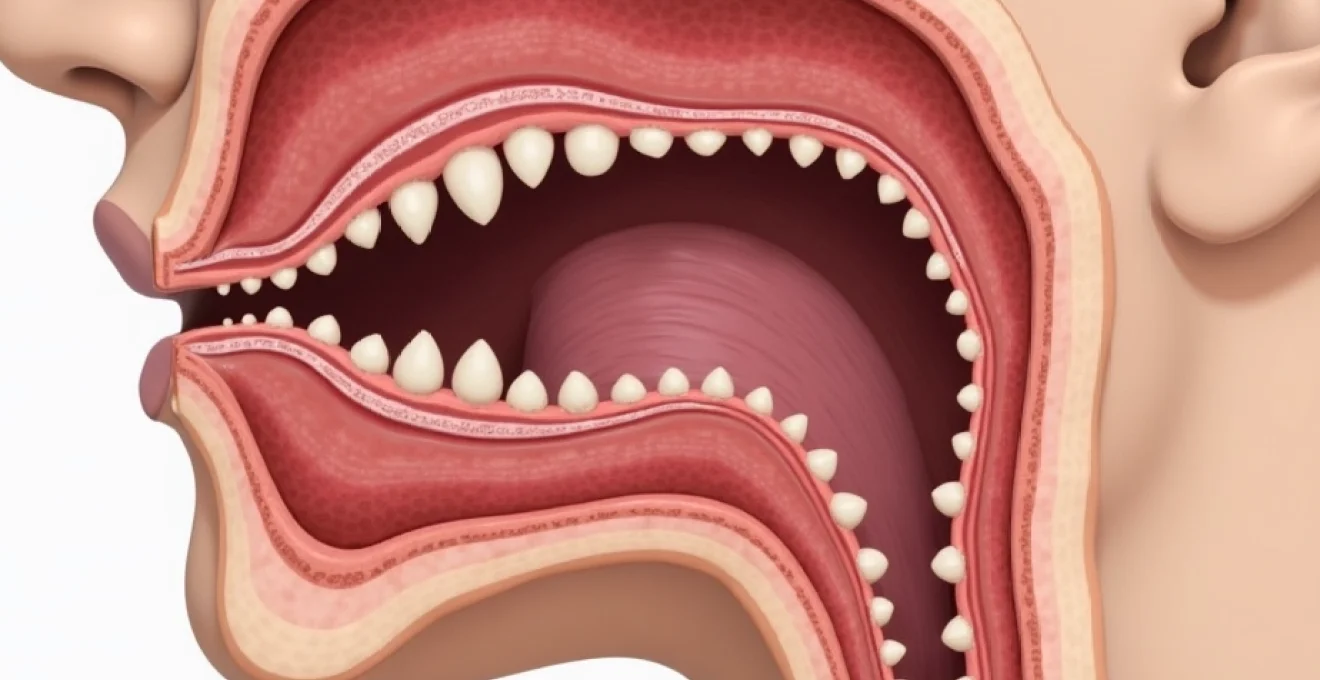
Anatomical structures and normal variations in posterior pharyngeal wall
The posterior pharynx contains numerous anatomical structures that naturally create holes, cavities, and depressions as part of their normal architecture. These features serve important physiological functions, particularly in immune surveillance and pathogen detection. Understanding these normal variations helps distinguish between benign anatomical features and potentially concerning pathological changes.
Palatine tonsil crypts and tonsillar fossa architecture
The palatine tonsils, situated on either side of the oropharynx, contain numerous crypts that appear as holes or openings on their surface. These tonsillar crypts are deep invaginations of the tonsillar epithelium that increase the surface area available for immune cell interaction with ingested antigens. The crypts can vary significantly in size and depth between individuals, with some appearing quite prominent and potentially concerning to patients who notice them for the first time.
These natural openings serve as sampling sites where the immune system encounters potential pathogens, allowing for early recognition and response to threats. However, the same crypts that provide immunological advantages can also become sites of debris accumulation, bacterial colonisation, and stone formation. Normal tonsil crypts typically appear pink and healthy, without surrounding inflammation or purulent discharge.
Lingual tonsil hypertrophy and lymphoid tissue distribution
The lingual tonsils, located at the base of the tongue, can also develop prominent follicular patterns and surface irregularities that may appear as holes or depressions. These structures are part of Waldeyer’s ring, a collection of lymphoid tissues that forms a protective barrier around the upper respiratory and digestive tract openings. Lingual tonsil hypertrophy can create noticeable surface variations that patients might interpret as abnormal cavities.
The distribution of lymphoid tissue throughout the pharynx creates a naturally irregular landscape with various peaks, valleys, and openings. This architectural complexity serves important immunological functions but can sometimes cause anxiety when patients discover these features during self-examination. Understanding the normal appearance of these structures helps differentiate physiological variations from pathological changes requiring medical attention.
Posterior pharyngeal wall follicular hyperplasia
Follicular hyperplasia of the posterior pharyngeal wall can create a cobblestone appearance with multiple small depressions and elevations. This condition often develops in response to chronic irritation from allergies, gastroesophageal reflux, or environmental pollutants. While not technically holes, these surface irregularities can appear concerning to patients unfamiliar with this benign condition.
The lymphoid follicles that create this appearance serve as immune surveillance posts, monitoring for potential threats in inhaled or ingested materials. When these follicles become hyperplastic due to chronic stimulation, they create a more pronounced surface texture that some patients might interpret as abnormal cavity formation. Recognition of this benign condition prevents unnecessary anxiety and inappropriate treatments.
Vallecular epiglottica and glossoepiglottic fold depressions
The valleculae epiglotticae are natural depressions located between the base of the tongue and the epiglottis, formed by the median and lateral glossoepiglottic folds. These anatomical spaces can appear as significant holes or cavities when viewed from certain angles, particularly during endoscopic examination or when patients use mirrors to examine their throat. These depressions serve important functions in swallowing mechanics and epiglottal movement.
During swallowing, these spaces help channel liquids around the epiglottis and into the oesophagus while protecting the airway. The size and prominence of these natural depressions vary considerably between individuals, with some appearing quite deep and potentially alarming to uninformed observers. Understanding their normal function and appearance prevents misinterpretation of these essential anatomical features as pathological conditions.
Infectious aetiologies causing pharyngeal cavity formation
Infections represent one of the most common causes of abnormal holes or cavities in the posterior pharynx. These infectious processes can create tissue breakdown, abscess formation, and chronic inflammatory changes that result in visible depressions or openings. The severity and appearance of these infectious cavities depend on the causative organisms, host immune response, and duration of the infectious process.
Peritonsillar abscess development and quinsy presentation
Peritonsillar abscesses, also known as quinsy, represent serious infections that can create significant tissue disruption and cavity formation in the peritonsillar space. These abscesses typically develop as complications of acute tonsillitis, when infection spreads beyond the tonsillar capsule into the surrounding connective tissue. The resulting inflammatory process can create visible depressions or openings as the abscess drains spontaneously or following medical intervention.
The clinical presentation of peritonsillar abscess includes severe throat pain, difficulty swallowing, fever, and characteristic changes in voice quality often described as a “hot potato” voice. The affected side typically shows marked swelling and displacement of surrounding structures, creating asymmetrical appearance of the throat. Early recognition and treatment of peritonsillar abscess are crucial to prevent complications such as airway obstruction or spread to deeper neck spaces.
Retropharyngeal space infections and deep neck abscesses
Retropharyngeal space infections can create serious cavities and tissue breakdown in the deep posterior pharynx. These infections are more common in young children due to the presence of retropharyngeal lymph nodes that typically regress with age. However, adults can develop retropharyngeal abscesses following trauma, foreign body ingestion, or as complications of other pharyngeal infections. The resulting tissue necrosis and abscess formation can create visible depressions or openings in the posterior pharyngeal wall.
The presentation of retropharyngeal space infection includes severe throat pain, fever, difficulty swallowing, and potential airway compromise. Patients may adopt unusual head positions to optimise airway patency, and the infection can rapidly progress to life-threatening complications if not promptly recognised and treated. The anatomical complexity of the deep neck spaces allows infections to spread along fascial planes, potentially reaching the mediastinum and causing thoracic complications.
Chronic tonsillitis with caseous debris accumulation
Chronic tonsillitis can lead to persistent inflammation and enlargement of tonsillar crypts, creating more prominent holes that accumulate caseous debris and bacterial biofilms. This condition often results from repeated episodes of acute tonsillitis that fail to completely resolve, leading to ongoing low-grade inflammation and structural changes in the tonsillar architecture. The enlarged crypts become more visible and may contain white or yellowish debris that patients often mistake for infection.
The caseous material found in these enlarged crypts consists of desquamated epithelial cells, food particles, bacteria, and inflammatory debris that becomes compacted over time. While often malodorous and aesthetically concerning, these accumulations are generally not dangerous unless they become secondarily infected. However, the persistent inflammatory state can contribute to recurrent throat symptoms and may warrant consideration of surgical intervention in severe cases.
Epstein-barr virus induced pharyngeal ulceration
Epstein-Barr virus (EBV) infection, commonly known as infectious mononucleosis, can cause significant pharyngeal changes including ulceration and tissue breakdown that may appear as holes or cavities. The virus has a particular affinity for lymphoid tissue, causing marked swelling and inflammation of tonsils and other pharyngeal lymphoid structures. In severe cases, the inflammatory process can lead to tissue necrosis and ulcer formation.
The pharyngeal manifestations of EBV infection typically include severe throat pain, marked tonsillar enlargement with exudative coating, and potential development of palatal petechiae. The tissue changes associated with severe EBV infection can create temporary depressions or openings that resolve as the acute infection subsides. Recognition of EBV-induced pharyngeal changes is important to avoid inappropriate antibiotic therapy and to monitor for potential complications such as airway obstruction from massive tonsillar enlargement.
Neoplastic conditions and malignant transformations
Malignant conditions affecting the oropharynx can present as holes, ulcerations, or cavitary lesions that require immediate medical attention. These neoplastic processes can arise from various cell types within the pharyngeal tissues and may present with subtle initial symptoms that gradually progress to more obvious structural changes. Early recognition of potentially malignant pharyngeal lesions is crucial for optimal treatment outcomes and patient survival.
Squamous cell carcinoma of oropharyngeal region
Squamous cell carcinoma represents the most common malignancy affecting the oropharynx and can present as ulcerative lesions that appear as holes or craters in the pharyngeal tissues. These malignant lesions typically develop gradually, initially appearing as small areas of tissue irregularity that progressively enlarge and ulcerate. The cancer cells destroy normal tissue architecture, creating characteristic excavated appearances that may be accompanied by surrounding inflammation and induration.
Risk factors for oropharyngeal squamous cell carcinoma include tobacco use, alcohol consumption, and human papillomavirus (HPV) infection. The clinical presentation often includes persistent throat pain, difficulty swallowing, referred ear pain, and potential voice changes. Any persistent pharyngeal lesion that fails to heal within two to three weeks warrants immediate medical evaluation and possible biopsy to rule out malignancy. Early detection significantly improves treatment outcomes and survival rates.
Hpv-related tonsillar malignancies and p16 positivity
Human papillomavirus-related oropharyngeal cancers have become increasingly common, particularly in younger patients without traditional risk factors such as tobacco and alcohol use. These HPV-positive malignancies often present as deep ulcerative lesions within the tonsillar crypts or base of tongue, creating cavitary appearances that may be initially mistaken for benign conditions. The viral transformation process can create characteristic tissue changes that experienced clinicians recognise as potentially malignant.
HPV-related oropharyngeal cancers typically demonstrate p16 protein overexpression, which serves as a surrogate marker for HPV involvement and has important prognostic implications. These tumours generally have better response rates to treatment and improved survival compared to HPV-negative cancers. However, the initial presentation can be subtle, with patients experiencing only mild throat discomfort or the sensation of something caught in their throat before more obvious symptoms develop.
Lymphoma presentation in waldeyer’s ring
Lymphomas affecting Waldeyer’s ring can present as rapidly enlarging masses that may ulcerate centrally, creating hole-like appearances in the affected tissues. Non-Hodgkin’s lymphomas are more common in this anatomical region, with diffuse large B-cell lymphoma being the most frequent subtype. These malignancies can cause dramatic tissue changes over relatively short periods, distinguishing them from more indolent processes.
The clinical presentation of pharyngeal lymphoma often includes rapid onset of throat pain, dysphagia, and potential airway compromise from mass effect. The tumours may outgrow their blood supply, leading to central necrosis and cavitation that creates characteristic crater-like appearances. Constitutional symptoms such as fever, night sweats, and unintentional weight loss may accompany the local pharyngeal changes, suggesting systemic lymphoma involvement.
Benign pharyngeal cysts and branchial cleft anomalies
Benign cystic lesions can develop within the pharyngeal tissues and may present as holes or depressions when they rupture spontaneously or following minor trauma. These cysts can arise from developmental anomalies, blocked ducts, or reactive inflammatory processes. Branchial cleft cysts, in particular, can present as pharyngeal lesions when they develop in atypical locations or communicate with the pharyngeal space through fistulous tracts.
The presentation of pharyngeal cysts varies depending on their size, location, and whether they have become infected or ruptured. Intact cysts typically appear as smooth, rounded swellings, while ruptured cysts may create irregular depressions or openings that can be concerning to patients. Infectious complications of pharyngeal cysts can cause surrounding inflammation and tissue changes that complicate the clinical picture and may require surgical intervention for definitive management.
Traumatic injuries and iatrogenic pharyngeal defects
Traumatic injuries to the pharynx can result in tissue disruption, perforation, and subsequent healing with scarring that creates visible depressions or openings. These injuries may result from penetrating trauma, blunt force injury, foreign body ingestion, or iatrogenic causes related to medical procedures. The severity and appearance of traumatic pharyngeal defects depend on the mechanism of injury, extent of tissue damage, and healing response of the affected individual.
Penetrating injuries to the pharynx can occur from various causes including motor vehicle accidents, assault, or accidental impalement with objects such as pens, utensils, or toys. Children are particularly susceptible to pharyngeal trauma from falls while carrying objects in their mouth or from playground accidents. The initial injury may appear minor externally but can cause significant internal damage to pharyngeal structures, blood vessels, and adjacent anatomical regions.
Iatrogenic pharyngeal injuries can result from medical procedures such as endotracheal intubation, endoscopy, tonsillectomy, or other surgical interventions in the head and neck region. These injuries may manifest as perforations, tissue defects, or areas of scarring that create visible depressions or openings. The healing process following traumatic or iatrogenic injury can result in contracture formation and tissue distortion that permanently alters the normal pharyngeal anatomy.
Foreign body ingestion represents another common cause of pharyngeal trauma, particularly in young children who commonly place objects in their mouth. Sharp or pointed objects can cause perforation or laceration of pharyngeal tissues, while larger objects may cause pressure necrosis if they become impacted. The resulting tissue damage can heal with scarring or chronic inflammation that creates visible depressions or openings that persist long after the initial injury.
Gastroesophageal reflux disease manifestations in pharynx
Gastroesophageal reflux disease (GERD) can cause significant changes to pharyngeal tissues through chronic exposure to acidic gastric contents. The posterior pharynx, larynx, and surrounding structures can develop inflammatory changes, ulceration, and subsequent scarring that may create visible depressions or tissue irregularities. Laryngopharyngeal reflux (LPR), a variant of GERD, specifically affects the upper respiratory tract and can cause characteristic tissue changes in the pharyngeal region.
The inflammatory effects of chronic acid exposure can lead to tissue breakdown, ulcer formation, and subsequent healing with fibrosis and scarring. These changes may create visible depressions or irregularities in the pharyngeal surface that patients might interpret as abnormal holes or cavities. The posterior pharynx and arytenoid regions are particularly susceptible to acid-induced injury due to their anatomical position and exposure patterns during reflux episodes.
Chronic GERD can also contribute to the development of other pharyngeal conditions such as follicular hyperplasia, chronic inflammation of lymphoid tissues, and increased susceptibility to infectious complications. The persistent inflammatory state created by acid exposure can alter the normal immune function of pharyngeal tissues and may contribute to the formation of more prominent crypts or surface irregularities that become visible during examination.
The diagnosis of GERD-related pharyngeal changes often requires correlation of symptoms with endoscopic findings and response to acid-suppression therapy. Patients may experience throat clearing, chronic c
ough, hoarseness, and a globus sensation (feeling of something stuck in the throat) that may persist despite treatment of the underlying reflux condition.
Treatment of GERD-related pharyngeal manifestations typically involves aggressive acid suppression with proton pump inhibitors, dietary modifications, and lifestyle changes to reduce reflux episodes. However, the tissue changes and scarring that develop from chronic acid exposure may persist even after successful treatment of the underlying reflux, creating permanent alterations in pharyngeal anatomy that can be concerning to patients and clinicians alike.
Diagnostic imaging protocols and endoscopic evaluation techniques
Accurate diagnosis of pharyngeal holes and cavities requires systematic evaluation using appropriate imaging modalities and endoscopic techniques. The choice of diagnostic approach depends on the clinical presentation, suspected aetiology, and accessibility of the lesion for direct visualization. Modern imaging technologies and advanced endoscopic equipment have significantly improved our ability to characterize pharyngeal abnormalities and guide appropriate treatment decisions.
Flexible fibreoptic laryngoscopy represents the gold standard for initial evaluation of pharyngeal lesions, allowing direct visualization of the entire pharyngeal and laryngeal anatomy. This office-based procedure provides excellent visualization of surface details, mucosal colour changes, and the three-dimensional relationships between anatomical structures. The procedure can be performed with topical anaesthesia and is generally well-tolerated by patients, making it an ideal screening tool for pharyngeal abnormalities.
Cross-sectional imaging studies, including computed tomography (CT) and magnetic resonance imaging (MRI), provide valuable information about deep tissue involvement, extent of lesions, and relationship to surrounding structures. CT imaging with intravenous contrast is particularly useful for evaluating infectious processes, characterizing mass lesions, and identifying potential complications such as abscess formation or vascular involvement. The ability to reconstruct images in multiple planes enhances the diagnostic capability and surgical planning when intervention is required.
Advanced endoscopic techniques such as narrow-band imaging (NBI) and chromoendoscopy can enhance the detection and characterization of subtle mucosal abnormalities that might be missed with conventional white light endoscopy. These specialized techniques highlight vascular patterns and surface architecture changes that may indicate early malignant transformation or chronic inflammatory processes. The enhanced contrast provided by these methods can be crucial for detecting pre-malignant changes in high-risk patients.
Ultrasound evaluation, while less commonly used for pharyngeal assessment, can provide valuable information about cystic lesions, lymph node involvement, and vascular structures in the neck. High-frequency ultrasound probes can characterize the internal architecture of pharyngeal masses and guide fine-needle aspiration procedures when tissue sampling is required. The real-time nature of ultrasound examination allows for dynamic assessment of swallowing function and structural movement during evaluation.
Biopsy techniques for pharyngeal lesions range from simple brush cytology for surface sampling to deep tissue biopsies obtained through various endoscopic approaches. The choice of biopsy method depends on the location, size, and suspected nature of the lesion. Office-based biopsies can be performed for easily accessible lesions, while deeper or more complex lesions may require surgical biopsy under general anaesthesia. Proper tissue handling and communication with pathologists are essential for accurate histopathological diagnosis.
Molecular diagnostic techniques, including HPV testing and p16 immunohistochemistry, have become standard components of the diagnostic workup for oropharyngeal malignancies. These tests provide crucial prognostic information and guide treatment decisions, particularly for squamous cell carcinomas of the oropharynx. The integration of molecular markers with traditional histopathological examination has revolutionized our understanding of pharyngeal malignancies and improved treatment outcomes.
Functional assessment of swallowing using videofluoroscopic swallow studies or fiberoptic endoscopic evaluation of swallowing (FEES) may be necessary when pharyngeal holes or cavities are associated with dysphagia or aspiration risks. These dynamic studies evaluate the coordination of swallowing mechanics and identify potential safety concerns that may influence treatment decisions. The combination of anatomical and functional assessment provides a comprehensive evaluation that guides optimal patient management strategies.