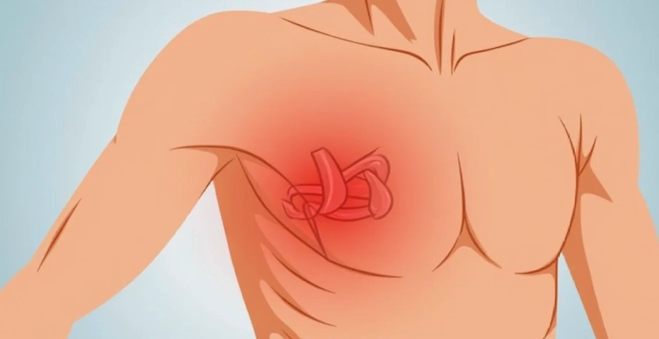
Left axillary pain, commonly referred to as pain under the left armpit, affects countless individuals and can stem from a diverse array of underlying conditions. The axillary region represents a complex anatomical area where multiple body systems converge, including muscular structures, lymphatic networks, neurological pathways, and dermatological tissues. Understanding the various causes of left armpit discomfort becomes crucial for proper self-assessment and determining when professional medical evaluation is necessary. From simple muscle strains to more complex systemic conditions, the spectrum of potential causes requires careful consideration and expert knowledge to navigate effectively.
Musculoskeletal causes of left axillary pain
The musculoskeletal system represents one of the most frequent sources of left axillary pain, with various muscle groups and connective tissues contributing to discomfort in this region. The intricate network of muscles surrounding the shoulder girdle and chest wall can become compromised through overuse, trauma, or repetitive stress patterns. Understanding these muscular causes helps differentiate between minor injuries and more serious underlying conditions that may require immediate attention.
Pectoralis major strain and microtrauma
The pectoralis major muscle, one of the largest muscles of the chest wall, extends from the sternum and clavicle to the upper arm bone, with its lateral fibres passing directly through the axillary region. Strain of this muscle commonly occurs during activities involving pushing, lifting, or sudden arm movements, particularly in individuals who engage in weightlifting, swimming, or contact sports. The resulting pain typically manifests as a deep, aching sensation that worsens with arm movement and may radiate into the armpit area.
Microtrauma to the pectoralis major can develop gradually through repetitive activities or sudden forceful contractions. This condition often affects individuals who perform repetitive overhead movements or those returning to physical activity after periods of inactivity. Recognition of early symptoms allows for prompt intervention and prevents progression to more severe muscle tears or chronic pain patterns.
Latissimus dorsi muscle tension and trigger points
The latissimus dorsi, often called the “lat” muscle, represents the broadest muscle of the back and plays a crucial role in arm adduction and extension. When this muscle develops tension or trigger points, pain can radiate anteriorly into the axillary region, creating discomfort that patients often describe as deep and persistent. Trigger points within the latissimus dorsi typically develop from poor posture, prolonged sitting, or repetitive pulling movements.
Professional athletes, particularly swimmers, rowers, and climbers, frequently experience latissimus dorsi-related axillary pain due to the repetitive nature of their training. The muscle’s extensive attachment points mean that dysfunction can create widespread pain patterns, often confusing both patients and healthcare providers about the true source of discomfort.
Intercostal muscle spasms and neuralgia
The intercostal muscles, situated between the ribs, play a vital role in respiratory function and trunk stability. When these muscles go into spasm or become inflamed, they can generate sharp, shooting pains that extend into the axillary region. Intercostal neuralgia, involving irritation of the nerves running between the ribs, creates particularly intense pain that may worsen with breathing, coughing, or sudden movements.
This condition often develops following respiratory infections, trauma to the chest wall, or as a complication of conditions such as shingles. The pain typically follows the path of the affected intercostal nerve, creating a band-like sensation that can extend from the back, around the side of the chest, and into the armpit area.
Serratus anterior dysfunction and scapular dyskinesis
The serratus anterior muscle, sometimes called the “boxer’s muscle,” originates from the upper ribs and inserts along the medial border of the scapula. Dysfunction of this muscle leads to altered scapular mechanics, often resulting in compensatory patterns that can generate axillary pain. Scapular dyskinesis , or abnormal movement of the shoulder blade, frequently accompanies serratus anterior weakness and can create persistent discomfort in the armpit region.
This muscle dysfunction commonly affects individuals with sedentary occupations who maintain prolonged forward head postures or those who perform repetitive overhead activities without adequate muscular balance. The resulting pain may be accompanied by visible winging of the shoulder blade and decreased shoulder function.
Lymphatic system pathologies in the left axilla
The axillary lymph nodes represent a critical component of the body’s immune system, filtering lymphatic fluid from the arm, breast, and upper chest wall. When these nodes become enlarged or inflamed, they can generate significant discomfort and concern for patients. Lymphatic causes of axillary pain range from benign reactive processes to more serious systemic conditions requiring immediate medical attention. Understanding the normal function and potential pathology of the lymphatic system helps distinguish between routine immune responses and conditions warranting further investigation.
Lymphadenopathy secondary to upper respiratory infections
Upper respiratory tract infections commonly cause reactive lymphadenopathy in the axillary region, as the lymphatic system responds to bacterial or viral pathogens. The axillary lymph nodes may become enlarged, tender, and palpable as they work to filter infectious agents from the lymphatic circulation. This reactive process typically develops within days of the onset of respiratory symptoms and may persist for several weeks following resolution of the primary infection.
The enlargement usually affects multiple lymph node groups, and patients may notice similar swelling in the neck or behind the ears. The nodes typically feel soft, mobile, and tender to touch, distinguishing them from more concerning fixed or hard lymphadenopathy. Monitoring the resolution of symptoms becomes important to ensure appropriate recovery and rule out persistent infectious processes.
Reactive lymph node hyperplasia from dermatological conditions
Various skin conditions affecting the arm, hand, or upper chest can trigger reactive lymph node hyperplasia in the axillary region. Conditions such as eczema, psoriasis, bacterial skin infections, or even minor cuts and abrasions can stimulate lymph node enlargement as part of the normal immune response. The degree of lymphadenopathy often correlates with the severity and extent of the dermatological condition.
Patients with chronic skin conditions may experience episodic axillary lymph node enlargement during disease flares. Understanding this connection helps patients recognise normal immune responses and avoid unnecessary anxiety about lymph node changes. However, persistent or progressively enlarging nodes warrant medical evaluation to rule out other underlying causes.
Axillary lymphangitis and cellulitis manifestations
Lymphangitis, or inflammation of the lymphatic vessels, can create visible red streaks extending from an infected area toward the axillary lymph nodes. This condition typically develops as a complication of bacterial skin infections, particularly those caused by Streptococcus or Staphylococcus species. The affected lymphatic vessels appear as red, warm, tender streaks on the skin surface, often accompanied by fever and malaise.
Cellulitis involving the axillary region can develop as an extension of lymphangitis or as a primary infectious process. The area becomes swollen, red, warm, and extremely tender, often with systemic symptoms including fever and chills. Prompt recognition and treatment of these conditions prevents progression to more serious complications such as abscess formation or sepsis.
Post-vaccination lymph node swelling and inflammatory response
Recent vaccination campaigns have highlighted the normal phenomenon of axillary lymphadenopathy following immunisation, particularly with mRNA vaccines. The lymph nodes on the same side as the vaccination site may become enlarged, tender, and palpable for several weeks following vaccination. This response indicates a normal immune system activation and antibody production.
While this reactive lymphadenopathy typically resolves spontaneously, patients should be aware of this potential side effect to avoid unnecessary concern. Healthcare providers must also consider recent vaccination history when evaluating patients with new axillary lymph node enlargement to provide appropriate reassurance and monitoring recommendations.
Dermatological and subcutaneous tissue disorders
The axillary region presents unique dermatological challenges due to its warm, moist environment and frequent friction from arm movement. The concentration of hair follicles, apocrine sweat glands, and sebaceous glands creates an ideal environment for various skin conditions that can generate significant pain and discomfort. Understanding common dermatological causes of axillary pain helps patients identify treatable conditions and implement appropriate preventive measures.
Hidradenitis suppurativa in the axillary region
Hidradenitis suppurativa represents a chronic inflammatory condition affecting the apocrine gland-bearing areas, with the axilla being one of the most commonly affected sites. This condition begins with the formation of painful nodules that may progress to form interconnected tunnels beneath the skin surface. The resulting pain can be severe and persistent, significantly impacting quality of life and daily activities.
The condition often follows a relapsing-remitting pattern, with periods of acute inflammation alternating with relative quiescence. Early recognition and treatment can prevent progression to more advanced stages characterised by extensive scarring and tissue destruction. The pain associated with hidradenitis suppurativa typically intensifies during flares and may be accompanied by purulent drainage and malodorous discharge.
Management requires a multidisciplinary approach combining medical therapy, lifestyle modifications, and sometimes surgical intervention. Understanding triggers such as stress, hormonal changes, and certain foods helps patients develop effective management strategies for this challenging condition.
Sebaceous cyst formation and secondary bacterial infection
Sebaceous cysts, also known as epidermoid cysts, commonly develop in the axillary region due to the high concentration of sebaceous glands in this area. These cysts form when keratin becomes trapped beneath the skin surface, creating a smooth, round mass that may be painless initially. However, when these cysts become infected or inflamed, they can generate significant pain, redness, and swelling.
Secondary bacterial infection of sebaceous cysts represents a common complication that requires prompt medical attention. The cyst becomes increasingly painful, warm to touch, and may develop purulent drainage.
Infected sebaceous cysts can rapidly progress to form abscesses if not properly treated, requiring surgical drainage and antibiotic therapy.
Understanding the signs of infection helps patients seek appropriate medical care before complications develop.
Contact dermatitis from antiperspirant aluminium compounds
Contact dermatitis affecting the axillary region frequently results from sensitisation to aluminium compounds found in antiperspirants and deodorants. This type IV hypersensitivity reaction can develop after years of use without problems, creating confusion for patients who have used the same products without previous issues. The resulting inflammation causes redness, itching, burning, and sometimes blistering of the affected skin.
Allergic contact dermatitis differs from irritant contact dermatitis in both mechanism and presentation. While irritant reactions typically develop immediately after exposure and affect anyone with sufficient contact, allergic reactions require prior sensitisation and affect only susceptible individuals. Identifying the specific allergen through patch testing allows for targeted avoidance and resolution of symptoms.
Folliculitis and pseudofolliculitis barbae
Folliculitis in the axillary region results from inflammation of hair follicles, often caused by bacterial infection, mechanical irritation, or occlusion. The condition presents as small, painful pustules surrounding hair follicles and can develop following shaving, tight clothing, or excessive sweating. Staphylococcus aureus represents the most common bacterial pathogen, though other organisms may be involved.
Pseudofolliculitis barbae, commonly called “razor bumps,” develops when curved hair shafts grow back into the skin after shaving, creating inflammatory papules and pustules. This condition disproportionately affects individuals with naturally curved hair and can become a chronic problem if not properly managed. The resulting inflammation can be quite painful and may lead to permanent scarring if severe.
Neurological conditions affecting left axillary sensation
Neurological causes of left axillary pain often present unique diagnostic challenges due to the complex innervation patterns of the axillary region. Multiple nerve pathways contribute to sensation in this area, including branches from the brachial plexus, intercostal nerves, and autonomic fibres. When these neural structures become compressed, inflamed, or damaged, they can generate various pain patterns ranging from sharp, shooting sensations to deep, burning discomfort that may be difficult to localise precisely.
The brachial plexus, a complex network of nerves emerging from the cervical and upper thoracic spine, provides motor and sensory innervation to the entire upper extremity. Compression or irritation of these nerve roots, often called thoracic outlet syndrome, can create pain that radiates into the axillary region. This condition frequently develops in individuals with specific anatomical variations, muscle imbalances, or occupations requiring prolonged overhead positioning. Understanding neurological pain patterns helps distinguish these conditions from muscular or vascular causes of axillary discomfort.
Intercostal neuralgia represents another significant neurological cause of axillary pain, occurring when the nerves running between the ribs become irritated or inflamed. This condition can develop following viral infections such as shingles, chest trauma, or as a complication of surgical procedures. The resulting pain typically follows the distribution of the affected nerve, creating a band-like sensation that extends from the spine, around the chest wall, and into the axillary region. Patients often describe this pain as sharp, electric, or burning in nature, with intensity that may vary throughout the day.
Peripheral neuropathy affecting the upper extremities can occasionally manifest with axillary pain, particularly in patients with diabetes, autoimmune conditions, or exposure to neurotoxic substances. The pain associated with neuropathy tends to be persistent, often described as burning or tingling, and may be accompanied by numbness or weakness in the affected distribution.
Neurological causes of axillary pain require careful evaluation to identify underlying conditions and prevent progression to permanent nerve damage.
Early recognition and appropriate treatment can often prevent long-term complications and improve patient outcomes.
Referred pain patterns and visceral causes
Referred pain represents a fascinating phenomenon where discomfort originates from internal organs or distant anatomical structures but is perceived in the axillary region. The complex interconnections of the nervous system allow pain signals from various sources to converge and create confusing symptom patterns that may mislead both patients and healthcare providers about the true underlying cause. Understanding these referral patterns becomes crucial for accurate diagnosis and appropriate treatment selection.
Cardiac conditions represent one of the most significant sources of referred axillary pain, particularly angina and myocardial infarction. The left axillary region commonly experiences pain during cardiac events due to shared neural pathways between the heart and this anatomical area. Patients experiencing cardiac-related axillary pain often describe it as pressure-like, squeezing, or heavy, frequently accompanied by chest discomfort, shortness of breath, nausea, or diaphoresis. Recognising cardiac referral patterns can be life-saving, as prompt medical attention during cardiac events significantly improves outcomes.
Pulmonary conditions such as pneumonia, pleuritis, or pulmonary embolism can also generate referred pain in the axillary region. The parietal pleura, which lines the chest cavity, shares innervation with superficial structures, allowing deep pulmonary inflammation to manifest as superficial pain. Patients with pleural inflammation often notice that their axillary pain worsens with deep inspiration, coughing, or position changes, helping distinguish pulmonary causes from other potential sources.
Gastrointestinal conditions, particularly those affecting the upper abdomen such as gallbladder disease or gastric ulceration, can occasionally refer pain to the left axillary region. The vagus nerve and sympathetic innervation of abdominal organs create complex referral patterns that may confuse the clinical picture. Patients with visceral pain referral often notice that their symptoms correlate with eating patterns, stress levels, or specific dietary triggers, providing important diagnostic clues for healthcare providers.
Spinal conditions affecting the cervical or upper thoracic regions frequently generate referred pain in the axillary area through nerve root irritation or compression. Herniated discs, spinal stenosis, or vertebral dysfunction can create pain that travels along nerve pathways into the armpit region. This type of referred pain often varies with neck position, arm movement, or spinal loading activities such as lifting or prolonged sitting.
Understanding the relationship between spinal health and axillary pain
helps prevent chronic pain development and maintains optimal shoulder function.
When to seek medical evaluation for persistent left axillary pain
Determining when left axillary pain requires professional medical evaluation can be challenging, particularly given the wide range of potential causes and varying symptom presentations. While many causes of axillary discomfort resolve spontaneously with conservative management, certain warning signs and symptom patterns warrant immediate medical attention to prevent serious complications or identify underlying conditions requiring specific treatment interventions.
Red flag symptoms that indicate the need for urgent medical evaluation include sudden onset of severe pain accompanied by chest discomfort, shortness of breath, or radiation to the jaw, neck, or left arm, as these may indicate cardiac events. Similarly, axillary pain associated with fever, rapidly spreading redness, red streaking extending from the armpit, or purulent drainage suggests serious infection requiring prompt antibiotic therapy. Any palpable masses or lumps in the axillary region that are hard, fixed, or progressively enlarging should be evaluated urgently to rule out malignant processes.
Persistent pain lasting longer than two weeks without improvement, despite appropriate rest and conservative measures, warrants medical assessment to identify underlying causes and prevent chronicity. Patients should also seek evaluation when axillary pain significantly interferes with daily activities, sleep patterns, or work performance, as effective treatment options are available for most conditions. Recurrent episodes of axillary pain, particularly when associated with skin changes, lymph node enlargement, or systemic symptoms, may indicate chronic conditions requiring ongoing management.
The presence of neurological symptoms such as numbness, tingling, weakness, or burning sensations extending beyond the axillary region suggests nerve involvement that may require specialised evaluation and treatment. Additionally, patients with risk factors for serious conditions, including personal or family history of cancer, cardiovascular disease, or autoimmune disorders, should maintain a lower threshold for seeking medical evaluation when experiencing new or changing axillary pain patterns.
Early medical evaluation of concerning axillary pain symptoms can prevent serious complications and ensure appropriate treatment selection for optimal outcomes.
When consulting healthcare providers about axillary pain, patients should prepare detailed information about symptom onset, duration, character, associated symptoms, and any triggering or relieving factors. This comprehensive history, combined with appropriate physical examination and diagnostic testing when indicated, allows healthcare providers to develop accurate diagnoses and effective treatment plans tailored to individual patient needs and underlying conditions.