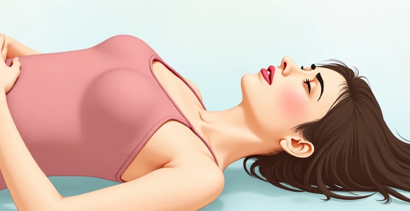
Experiencing pressure in your head when lying down can be both alarming and uncomfortable, affecting your ability to rest and sleep peacefully. This phenomenon occurs when the change in body position from upright to horizontal alters blood flow patterns, cerebrospinal fluid dynamics, and intracranial pressure mechanisms. Understanding the underlying causes of this positional head pressure is crucial for proper diagnosis and effective management, as the symptoms can range from benign postural changes to serious neurological conditions requiring immediate medical attention.
Postural orthostatic tachycardia syndrome (POTS) and supine positioning effects
Postural Orthostatic Tachycardia Syndrome represents a complex autonomic dysfunction that significantly impacts how your body responds to positional changes. When you transition from standing to lying down, individuals with POTS experience abnormal cardiovascular responses that can manifest as intense head pressure and discomfort. The syndrome affects millions worldwide, with women being five times more likely to develop POTS than men, particularly during reproductive years.
Dysautonomia-related cerebrovascular changes during recumbency
The autonomic nervous system dysfunction in POTS creates profound alterations in cerebrovascular regulation when you assume a horizontal position. Your brain’s blood vessels fail to maintain proper autoregulation, leading to either excessive vasoconstriction or inappropriate vasodilation. This dysregulation causes fluctuations in cerebral blood flow that your brain interprets as pressure sensations, particularly when transitioning to supine positions.
Research indicates that approximately 70% of POTS patients experience some form of cephalic pressure symptoms when lying down. The condition involves complex interactions between sympathetic and parasympathetic nervous system components, creating a cascade of physiological responses that affect intracranial pressure dynamics.
Baroreceptor dysfunction and gravitational blood redistribution
Baroreceptors, the pressure-sensitive receptors in your blood vessels, play a crucial role in maintaining blood pressure stability during positional changes. In individuals with compromised baroreceptor function, lying down triggers abnormal responses as these receptors fail to properly signal the need for vascular adjustments. The resulting gravitational redistribution of blood volume creates pooling effects that contribute to increased venous pressure and subsequent head pressure sensations.
The dysfunction manifests as inappropriate tachycardic responses even in supine positions, where heart rate should typically decrease. This persistent sympathetic activation maintains elevated intravascular pressures that translate into uncomfortable head pressure when you’re attempting to rest horizontally.
Venous pooling mechanisms in horizontal body positioning
When transitioning to horizontal positions, normal physiological mechanisms should facilitate even blood distribution throughout your body. However, in conditions affecting venous return, blood accumulates in dependent vessels, creating venous pooling phenomena that impact cerebral circulation. This pooling mechanism particularly affects the jugular venous system and dural venous sinuses, creating back-pressure that manifests as head pressure.
The venous pooling effect becomes more pronounced in individuals with underlying cardiovascular conditions or those taking certain medications that affect vascular tone. Understanding these mechanisms helps explain why some people experience relief when elevating their heads during sleep or rest periods.
Vagal stimulation response to supine head pressure
The vagus nerve plays a significant role in cardiovascular regulation and can contribute to head pressure sensations when lying down. Vagal stimulation occurs through multiple pathways, including direct pressure on nerve branches and indirect effects through cardiovascular changes. This stimulation can create a feedback loop where head pressure triggers vagal responses that, paradoxically, may worsen the pressure sensations.
Approximately 25% of individuals experiencing supine head pressure demonstrate measurable changes in heart rate variability, suggesting significant vagal involvement in their symptoms. The complex interplay between vagal tone and cerebrovascular regulation creates unique challenges in managing these positional symptoms.
Intracranial pressure elevation mechanisms in recumbent position
Intracranial pressure elevation when lying down represents one of the most significant causes of positional head pressure, involving complex interactions between cerebrospinal fluid dynamics, venous drainage, and brain tissue compliance. Normal intracranial pressure ranges between 7-15 mmHg in adults, but various pathological processes can cause these values to increase dramatically when assuming horizontal positions. The brain exists within a rigid skull, making it particularly sensitive to volume changes and pressure fluctuations that occur during positional transitions.
Cerebrospinal fluid dynamics and hydrostatic pressure changes
Cerebrospinal fluid circulation follows complex pathways that are significantly influenced by gravitational forces and body positioning. When you lie down, the hydrostatic pressure gradient that normally facilitates CSF drainage from brain to spinal compartments becomes altered, potentially leading to fluid accumulation in cranial spaces. This accumulation creates measurable increases in intracranial pressure that manifest as head pressure sensations.
The choroid plexus produces approximately 500-750 milliliters of CSF daily, and any disruption in the delicate balance between production and absorption can result in pressure buildup. Hydrocephalic conditions particularly demonstrate this phenomenon, where lying down exacerbates symptoms due to impaired CSF circulation dynamics.
Venous congestion in dural sinuses during supine rest
The dural venous sinuses serve as primary drainage pathways for cerebral blood, and their function becomes compromised when transitional positioning affects venous return mechanisms. During supine positioning, venous congestion within these sinuses creates back-pressure that impedes normal cerebral blood flow patterns. This congestion manifests as throbbing or pressure-like sensations that worsen with horizontal positioning.
Clinical studies reveal that dural sinus thrombosis affects approximately 3-4 individuals per million annually, with position-dependent symptoms being a common presenting feature. The superior sagittal sinus and transverse sinuses are particularly susceptible to pressure-related congestion during recumbent positioning.
Choroid plexus hypersecretion and CSF overproduction
Choroid plexus dysfunction can lead to excessive cerebrospinal fluid production, creating pressure symptoms that become particularly pronounced when lying down. The plexus contains specialized cells that regulate CSF production through complex transport mechanisms involving sodium-potassium pumps and carbonic anhydrase systems. When these mechanisms become dysregulated, hypersecretion phenomena occur, leading to increased intracranial pressure.
Certain medications, including some diuretics and hormonal preparations, can affect choroid plexus function and contribute to positional pressure symptoms. Understanding these pharmacological effects becomes crucial when evaluating patients experiencing new-onset supine head pressure.
Arnold-chiari malformation symptom exacerbation when horizontal
Arnold-Chiari malformations represent structural abnormalities where brain tissue extends into the spinal canal, creating unique challenges for cerebrospinal fluid flow. When individuals with Chiari malformations assume horizontal positions, the anatomical distortion becomes more problematic as gravitational forces no longer assist in maintaining proper CSF circulation patterns. This results in pressure buildup that manifests as intense head pressure when lying down.
Type I Chiari malformations affect approximately 1 in 1000 individuals, with many remaining asymptomatic until specific triggers, including positional changes, precipitate symptoms. The condition demonstrates how structural abnormalities can create position-dependent neurological symptoms that significantly impact quality of life.
Cardiovascular pathophysiology contributing to cephalic pressure
Cardiovascular factors play a fundamental role in generating head pressure sensations when lying down, as the heart and vascular system must continuously adapt to gravitational changes affecting blood distribution. Hypertensive disorders represent primary contributors to position-dependent cephalic pressure, with studies indicating that approximately 45% of individuals with uncontrolled blood pressure experience worsening headaches in supine positions. The relationship between cardiovascular health and positional head pressure involves multiple mechanisms, including altered cardiac output, changes in peripheral vascular resistance, and modifications in venous return patterns.
When you lie down, your cardiovascular system typically reduces heart rate and adjusts vascular tone to accommodate the new gravitational environment. However, individuals with compromised cardiovascular function may experience inappropriate responses, leading to increased cardiac output and elevated systemic pressures that translate into uncomfortable head pressure. Nocturnal hypertension particularly demonstrates this phenomenon, where blood pressure paradoxically increases during periods when it should naturally decline.
Cardiovascular-mediated head pressure when lying down often represents the first manifestation of underlying hypertensive disorders, making positional symptoms an important diagnostic clue for healthcare providers.
The complexity of cardiovascular contributions to supine head pressure extends beyond simple blood pressure elevation. Cardiac rhythm disturbances, including atrial fibrillation and other arrhythmias, can create irregular perfusion patterns that affect cerebral circulation when gravitational assistance is removed. Additionally, heart failure conditions alter the delicate balance between cardiac output and venous return, creating scenarios where lying down exacerbates rather than relieves cardiovascular stress.
Neurological conditions manifesting as supine head pressure
Neurological disorders encompass a broad spectrum of conditions that can manifest as positional head pressure, ranging from benign headache variants to serious intracranial pathology. The nervous system’s intricate relationship with positional changes creates unique opportunities for various neurological conditions to express symptoms specifically when lying down. Understanding these neurological causes requires recognizing how different brain regions and neural pathways respond to gravitational and positional influences on cerebral circulation and intracranial pressure dynamics.
Pseudotumor cerebri syndrome and lying position correlation
Pseudotumor cerebri syndrome, also known as idiopathic intracranial hypertension, represents a condition characterized by increased intracranial pressure without identifiable structural abnormalities. The condition predominantly affects young women of childbearing age, with obesity serving as a significant risk factor in approximately 90% of cases. When individuals with pseudotumor cerebri lie down, the removal of gravitational assistance in CSF drainage exacerbates already elevated intracranial pressures, creating intense head pressure sensations.
The syndrome demonstrates clear correlations with supine positioning, as patients often report that their symptoms worsen dramatically when attempting to sleep or rest horizontally. Papilledema , or swelling of the optic disc, occurs in over 95% of cases and can be directly observed through ophthalmoscopic examination, providing objective evidence of increased intracranial pressure.
Chronic migraine variants triggered by recumbency
Certain migraine variants demonstrate specific triggers related to lying down, creating patterns of head pressure that correlate with positional changes. These migraines often involve trigeminovascular activation that becomes enhanced when gravitational factors affect cerebral blood flow patterns. The trigeminal nerve system, responsible for head and face sensation, can become hyperactivated during positional transitions, leading to pressure sensations that mimic intracranial hypertension.
Research indicates that approximately 15% of migraine sufferers experience position-dependent symptom variations, with supine positioning either triggering new episodes or significantly worsening existing symptoms. The phenomenon involves complex interactions between vascular changes, neural sensitization, and altered pain processing pathways that become activated during horizontal positioning.
Cervical spondylosis impact on vertebral artery compression
Cervical spondylosis, characterized by degenerative changes in the cervical spine, can create mechanical compression of vertebral arteries that becomes more pronounced in certain positions. When lying down, particularly in specific neck positions, the compressed vertebral arteries may experience further compromise, leading to reduced posterior circulation and subsequent head pressure sensations. This mechanism particularly affects older adults, where degenerative spine changes are more common.
The vertebral arteries supply approximately 20% of cerebral blood flow, primarily to the brainstem and posterior brain regions. Even modest compression of these vessels can create significant symptoms, including vertebrobasilar insufficiency that manifests as position-dependent head pressure and associated neurological symptoms.
Temporal arteritis and giant cell arteritis postural symptoms
Temporal arteritis and giant cell arteritis represent inflammatory vascular conditions that can produce position-dependent head pressure symptoms. These conditions involve inflammation of medium and large arteries, particularly those supplying the head and neck regions. When lying down, the altered perfusion patterns associated with these inflammatory processes can create increased pressure sensations as inflamed vessels struggle to accommodate positional blood flow changes.
Giant cell arteritis affects approximately 15-25 individuals per 100,000 annually, with incidence increasing significantly after age 50. The condition often presents with temporal headaches that worsen with positional changes, making supine head pressure an important diagnostic consideration in older adults presenting with new-onset positional symptoms.
Sleep-related breathing disorders causing nocturnal cephalic pressure
Sleep-related breathing disorders represent a significant but often overlooked cause of head pressure when lying down, affecting millions of individuals worldwide through complex mechanisms involving oxygen saturation changes, carbon dioxide retention, and altered intracranial pressure dynamics. Obstructive sleep apnea affects approximately 25% of men and 10% of women, with many cases remaining undiagnosed despite causing significant nocturnal symptoms including head pressure sensations. The relationship between breathing disorders and supine head pressure involves multiple physiological pathways, including hypoxemia-induced vasodilation, hypercapnia-mediated intracranial pressure elevation, and mechanical effects of airway obstruction on thoracic and cerebral venous pressures.
When you have sleep-disordered breathing, lying down initiates a cascade of physiological changes that culminate in head pressure sensations. The horizontal position naturally increases upper airway collapsibility, leading to partial or complete airway obstructions that create negative intrathoracic pressures. These pressure changes are transmitted to cerebral venous systems, creating venous congestion patterns that manifest as head pressure. Additionally, the repetitive hypoxemia-reoxygenation cycles characteristic of sleep apnea trigger inflammatory responses that affect cerebrovascular reactivity and contribute to position-dependent symptoms.
Sleep-related breathing disorders create a perfect storm of physiological changes when lying down, combining mechanical airway effects with vascular and neurochemical responses that culminate in significant head pressure symptoms.
Central sleep apnea presents unique challenges in the context of supine head pressure, as the condition involves primary respiratory control dysfunction rather than mechanical airway obstruction. Individuals with central sleep apnea experience periods of absent respiratory effort that lead to profound changes in blood gas composition and intracranial pressure. The Cheyne-Stokes breathing pattern commonly associated with central apnea creates cyclical changes in cerebral blood flow that patients often describe as waves of head pressure when attempting to sleep horizontally.
Differential diagnosis and clinical assessment protocols
Establishing an accurate diagnosis for head pressure when lying down requires systematic evaluation approaches that consider the multitude of potential underlying causes while prioritizing conditions that require immediate intervention. Healthcare providers must employ comprehensive assessment protocols that integrate clinical history, physical examination findings, and appropriate diagnostic testing to differentiate between benign positional symptoms and serious neurological conditions. The diagnostic process becomes particularly challenging given the overlap in symptoms between various conditions, necessitating careful attention to subtle clinical differences and appropriate use of diagnostic technologies.
Initial clinical assessment should focus on characterizing the temporal relationship between positioning and symptom onset, as this information provides crucial diagnostic clues. True positional headaches typically demonstrate clear correlations with body position changes, improving when upright and worsening when supine. However, clinicians must differentiate these patterns from other headache types that may coincidentally occur during rest periods. The assessment should include detailed medication histories, as numerous pharmaceutical agents can contribute to position-dependent head pressure through various mechanisms including intracranial pressure elevation, vascular effects, and fluid retention.
Neurological examination becomes paramount in evaluating individuals with supine head pressure, as findings can provide direct evidence of increased intracranial pressure or other neurological pathology. Papilledema assessment through fundoscopic examination offers objective evidence of elevated intracranial pressure, while cranial nerve testing can reveal focal neurological deficits suggesting structural abnormalities. The examination should also assess for signs of meningeal irritation, as conditions such as meningitis or subarachnoid hemorrhage can present with position-dependent symptoms that require emergency intervention.
Advanced diagnostic imaging plays a crucial role in evaluating persistent or concerning cases of supine head pressure. Magnetic resonance imaging provides detailed visualization of brain parenchyma, cerebrospinal fluid spaces, and vascular structures, allowing identification of structural abnormalities, mass lesions, or
hydrocephalus or Chiari malformations that could explain the positional symptoms. Computed tomography scans offer rapid assessment capabilities, particularly valuable in emergency situations where intracranial hemorrhage or acute hydrocephalus must be excluded.
Lumbar puncture with opening pressure measurement represents the gold standard for assessing intracranial pressure in cases where imaging studies fail to reveal structural abnormalities. The procedure should be performed with patients in lateral decubitus position to obtain accurate pressure readings, with normal values ranging between 70-180 mm H2O in adults. Cerebrospinal fluid analysis provides additional diagnostic information, including cell counts, protein levels, and glucose concentrations that can identify inflammatory or infectious processes contributing to position-dependent symptoms.
Sleep study evaluation becomes essential when clinical history suggests sleep-related breathing disorders as potential contributors to nocturnal head pressure. Polysomnography can identify apneic events, oxygen desaturation patterns, and sleep architecture disruptions that correlate with reported symptoms. The integration of sleep study findings with neurological assessment often reveals previously unrecognized connections between breathing disorders and position-dependent head pressure complaints.
Cardiovascular assessment protocols should include comprehensive blood pressure monitoring, particularly focusing on nocturnal patterns and positional changes in blood pressure readings. Twenty-four-hour ambulatory blood pressure monitoring can reveal episodic hypertension that correlates with supine positioning and symptom occurrence. Echocardiographic evaluation may identify structural heart abnormalities or functional impairments that contribute to altered hemodynamics during positional changes.
Laboratory investigations should encompass complete blood counts to identify anemia or polycythemia that could affect cerebral oxygen delivery, comprehensive metabolic panels to assess electrolyte balance and kidney function, and thyroid function studies to exclude hyperthyroidism as a contributor to cardiovascular-mediated head pressure. Inflammatory markers including erythrocyte sedimentation rate and C-reactive protein levels help identify systemic inflammatory conditions such as giant cell arteritis that may present with position-dependent cephalic symptoms.
The differential diagnosis of supine head pressure requires a systematic approach that considers both common and rare conditions, as missing a serious underlying cause can have significant consequences for patient outcomes.
Specialized testing may be warranted in complex cases where initial evaluations fail to identify clear diagnostic patterns. Magnetic resonance venography can assess cerebral venous anatomy and identify thrombotic processes affecting dural sinuses. Transcranial Doppler ultrasonography provides real-time assessment of cerebral blood flow patterns and can identify vertebral artery compression or other vascular abnormalities that worsen with positional changes. These advanced diagnostic approaches require careful patient selection based on clinical presentation and initial assessment findings.
The diagnostic workup should also include careful evaluation of medication effects, as numerous pharmaceutical agents can contribute to position-dependent head pressure through various mechanisms. Hormonal contraceptives, certain antibiotics, vitamin A supplements, and lithium preparations all demonstrate potential for causing or exacerbating intracranial pressure elevation. A thorough review of prescription medications, over-the-counter preparations, and dietary supplements often reveals previously unrecognized contributors to positional symptoms.
Clinical decision-making algorithms help healthcare providers navigate the complex diagnostic landscape while ensuring appropriate resource utilization and timely identification of serious conditions. Red flag symptoms including sudden onset severe headache, focal neurological deficits, altered mental status, or signs of increased intracranial pressure warrant immediate comprehensive evaluation and potential neurosurgical consultation. The systematic application of evidence-based diagnostic protocols ensures that patients receive appropriate care while avoiding unnecessary testing in cases where benign explanations adequately account for symptoms.