Facial asymmetry during speech represents a fascinating intersection of neurology, anatomy, and human communication. While many people assume that speaking involves symmetrical facial movements, research reveals that approximately 76% of individuals naturally exhibit greater mouth opening on the right side during spontaneous speech. This phenomenon reflects the complex neural pathways that control facial expression and language production, highlighting the intricate relationship between brain hemispheres and motor control.
When facial asymmetry becomes pronounced or develops suddenly, it often signals underlying medical conditions that require immediate attention. The causes range from temporary inflammatory conditions to serious neurological disorders, each presenting unique challenges for diagnosis and treatment. Understanding these various aetiologies helps distinguish between normal speech patterns and pathological conditions that demand medical intervention.
Neurological disorders affecting unilateral facial motor control
The seventh cranial nerve, also known as the facial nerve, plays a crucial role in controlling the muscles responsible for facial expression and speech articulation. When this nerve becomes compromised through various neurological conditions, the result is often noticeable facial asymmetry that becomes particularly apparent during speaking. These disorders can affect the nerve at different anatomical locations, from its origin in the brainstem to its peripheral branches in the face.
Bell’s palsy and idiopathic facial nerve paralysis
Bell’s palsy represents the most common cause of acute unilateral facial weakness, affecting approximately 15 to 30 people per 100,000 annually. This condition results from inflammation and swelling of the seventh cranial nerve, leading to temporary facial paralysis that typically affects one side of the face. The characteristic drooping of the mouth corner makes speaking particularly challenging, as patients struggle to articulate words clearly and may experience drooling during conversation.
The onset of Bell’s palsy is typically sudden, with symptoms reaching peak severity within 48 to 72 hours. During this acute phase, patients often report difficulty with basic facial expressions such as smiling, frowning, or closing the affected eyelid. Speech production becomes notably asymmetrical , with the paralysed side of the mouth failing to participate in normal articulation patterns. This creates a distinctive lopsided appearance during conversation that can be both functionally limiting and emotionally distressing for patients.
Viral infections, particularly herpes simplex virus, varicella-zoster virus, and Epstein-Barr virus, are suspected triggers for Bell’s palsy, though the exact mechanism remains unclear. The condition shows a slight female predominance and tends to affect individuals between ages 15 and 60 most frequently. Pregnancy, diabetes, and upper respiratory infections may increase the risk of developing this condition.
Stroke-related facial weakness and upper motor neurone lesions
Cerebrovascular accidents affecting the motor cortex or internal capsule can result in facial weakness that differs significantly from peripheral nerve palsies. Unlike Bell’s palsy, which affects both upper and lower facial muscles, strokes typically spare the forehead muscles due to bilateral cortical innervation of the upper facial region. This creates a characteristic pattern where patients can wrinkle their foreheads but cannot smile symmetrically or control the lower portion of their face effectively.
The speech impairment associated with stroke-related facial weakness often accompanies other neurological deficits such as dysarthria, aphasia, or limb weakness. Central facial paralysis presents unique challenges because it frequently occurs alongside cognitive and language processing difficulties, making rehabilitation more complex than in cases of isolated peripheral nerve damage.
Emergency recognition of stroke symptoms is critical, as the phrase “time is brain” emphasises the importance of rapid medical intervention. The FAST assessment (Face drooping, Arm weakness, Speech difficulties, Time to call emergency services) includes facial asymmetry as a key diagnostic criterion, highlighting the significance of unilateral mouth involvement in identifying serious cerebrovascular events.
Ramsay hunt syndrome and herpes zoster oticus
Ramsay Hunt syndrome occurs when the varicella-zoster virus reactivates and affects the geniculate ganglion of the seventh cranial nerve. This condition combines the facial paralysis characteristic of Bell’s palsy with the additional features of vesicular rash in the ear canal and severe otalgia. The facial weakness in Ramsay Hunt syndrome often proves more severe and has a poorer prognosis for recovery compared to idiopathic Bell’s palsy.
Patients with Ramsay Hunt syndrome frequently experience hearing loss, tinnitus, and vertigo alongside their facial paralysis. The speech difficulties extend beyond simple motor weakness to include challenges with auditory processing and balance maintenance during conversation. Early antiviral treatment combined with corticosteroids offers the best chance for facial function recovery, though complete restoration occurs in only about 70% of cases.
Acoustic neuroma and cerebellopontine angle tumours
Benign tumours arising from the eighth cranial nerve, known as acoustic neuromas or vestibular schwannomas, can compress adjacent structures including the facial nerve as they grow. These slow-growing masses typically present with gradual hearing loss and tinnitus before facial weakness becomes apparent. When facial involvement occurs, it usually indicates significant tumour size and potential compression of multiple cranial nerves within the cerebellopontine angle.
The facial weakness associated with acoustic neuromas develops insidiously, often progressing from subtle asymmetry during animated speech to more pronounced paralysis over months or years. Unlike acute inflammatory conditions, the gradual onset allows for some compensatory mechanisms to develop, though complete functional recovery depends on timely surgical intervention and the extent of nerve involvement at diagnosis.
Multiple sclerosis and demyelinating facial neuropathy
Multiple sclerosis can affect the facial nerve through demyelinating lesions within the brainstem, resulting in episodes of facial weakness that may recur or become permanent. The unpredictable nature of MS means that facial symptoms can appear as isolated events or as part of broader neurological relapses affecting multiple body systems simultaneously.
Distinguishing MS-related facial weakness from other causes requires careful neurological evaluation and often magnetic resonance imaging to identify characteristic white matter lesions. The speech difficulties in MS patients extend beyond mechanical facial weakness to include dysarthria related to cerebellar involvement and cognitive processing changes that affect language production and comprehension.
Traumatic injuries and surgical complications
Physical trauma and surgical procedures represent significant causes of facial nerve injury, often resulting in immediate and severe functional deficits. These iatrogenic and traumatic injuries present unique challenges because they frequently involve complete nerve transection or severe crushing injuries that may require surgical repair for optimal recovery. The mechanism and location of injury significantly influence both the initial presentation and long-term prognosis for facial function restoration.
Temporal bone fractures and facial nerve transection
High-energy trauma affecting the temporal bone can result in facial nerve injury through either longitudinal or transverse fracture patterns. Longitudinal fractures, which account for approximately 80% of temporal bone injuries, typically cause less severe facial nerve damage compared to transverse fractures that directly cross the nerve’s course through the fallopian canal. The immediate assessment of facial function following head trauma is crucial for determining the need for urgent surgical intervention.
Complete facial nerve transection from temporal bone fractures results in immediate and total facial paralysis on the affected side. The inability to close the eye properly creates additional risks for corneal damage and vision loss, requiring immediate protective measures and ophthalmological consultation. Speech becomes significantly impaired due to the loss of labial seal and precise tongue coordination with facial muscles.
The assessment of facial nerve function in trauma patients requires careful examination once the patient is medically stable, as delayed recovery may indicate the need for surgical exploration and nerve grafting procedures.
Parotidectomy-related facial nerve damage
Surgical removal of the parotid gland, whether for benign or malignant conditions, carries inherent risks to the facial nerve due to its intimate anatomical relationship with the gland. The main trunk of the facial nerve divides into upper and lower divisions within the parotid gland, making nerve preservation technically challenging during tumour excision. Modern surgical techniques emphasise nerve monitoring and careful dissection to minimise functional deficits.
Post-parotidectomy facial weakness can range from temporary neuropraxia to permanent paralysis depending on the extent of surgical manipulation and tumour involvement. Patients often experience varying degrees of facial asymmetry during the recovery period, with speech patterns showing gradual improvement as nerve function returns. The psychological impact of facial disfigurement following parotidectomy requires comprehensive rehabilitation approaches that address both functional and aesthetic concerns.
Middle ear surgery and iatrogenic nerve injury
Otological procedures such as mastoidectomy, tympanoplasty, and stapedectomy pose risks to the facial nerve due to its course through the middle ear space. The tympanic segment of the facial nerve lies in close proximity to surgical landmarks, making inadvertent injury possible even with experienced surgeons. The use of facial nerve monitoring during these procedures has significantly reduced the incidence of iatrogenic facial paralysis.
When facial nerve injury occurs during middle ear surgery, the functional deficit may be temporary or permanent depending on the mechanism and severity of damage. Immediate recognition of facial weakness in the postoperative period allows for prompt intervention and optimises the chances for functional recovery through medical management or surgical repair.
Penetrating facial trauma and peripheral nerve laceration
Sharp force injuries to the face, including knife wounds, glass lacerations, and projectile injuries, can cause direct damage to peripheral branches of the facial nerve. The anatomical distribution of weakness provides clues to the specific nerve branches affected, with injuries anterior to a vertical line through the lateral canthus typically requiring surgical exploration and repair.
The timing of surgical repair for traumatic facial nerve lacerations significantly influences functional outcomes. Primary repair within 72 hours of injury generally produces the best results, while delayed reconstruction may require nerve grafting procedures with less predictable functional recovery. The speech impairments following peripheral facial nerve trauma often show good recovery potential with appropriate surgical management and rehabilitation.
Congenital abnormalities and developmental disorders
Congenital facial paralysis represents a diverse group of conditions that affect facial motor function from birth, resulting in lifelong challenges with speech articulation, facial expression, and social communication. These conditions range from isolated facial nerve anomalies to complex syndromic presentations involving multiple cranial nerves and developmental structures. The impact on speech development is particularly significant because infants learn to articulate sounds through visual and proprioceptive feedback from their own facial movements.
Möbius syndrome exemplifies the most recognised congenital cause of bilateral facial paralysis, typically involving cranial nerves VI and VII with variable involvement of other cranial nerves. Children with Möbius syndrome face unique challenges in developing speech patterns because they cannot observe their own facial movements or receive normal sensory feedback from muscle contractions. The absence of facial expression also affects their ability to engage in non-verbal communication, requiring specialised speech therapy approaches that emphasise alternative communication strategies.
Hemifacial microsomia and other craniofacial malformation syndromes can involve unilateral facial nerve hypoplasia or absence, resulting in asymmetrical facial development and function. These conditions often require multidisciplinary management involving plastic surgeons, orthodontists, and speech therapists to optimise both functional and aesthetic outcomes. The timing of surgical interventions must be carefully coordinated with speech development milestones to maximise therapeutic benefits.
Congenital facial nerve paralysis may also occur as an isolated finding without associated syndromes, possibly resulting from intrauterine positioning, birth trauma, or developmental anomalies of the facial nerve nucleus. Early intervention with facial reanimation procedures, such as nerve transfers or muscle transpositions, can significantly improve functional outcomes and speech development when performed during childhood.
The management of congenital facial paralysis requires a comprehensive understanding of normal speech development patterns and the specific challenges faced by children who cannot achieve symmetrical facial movements during communication.
Inflammatory and autoimmune conditions
Various inflammatory and autoimmune disorders can target the facial nerve, resulting in acute or chronic facial paralysis with associated speech difficulties. These conditions often present diagnostic challenges because they may mimic more common causes of facial weakness while requiring specific immunosuppressive treatments for optimal management. The inflammatory process can affect the nerve at multiple levels, from the brainstem nucleus to the peripheral nerve branches, depending on the underlying pathology.
Sarcoidosis frequently affects the nervous system, with facial nerve involvement being one of the most common neurological manifestations of this multisystem granulomatous disease. Unlike Bell’s palsy, sarcoid-related facial weakness may be bilateral and can be associated with other neurological symptoms such as hearing loss, vestibular dysfunction, or cognitive changes. The chronic nature of sarcoidosis means that facial weakness may persist or recur without appropriate immunosuppressive treatment.
Lyme disease, caused by Borrelia burgdorferi infection, can produce facial nerve palsy as part of the early disseminated phase of infection. The facial weakness in Lyme disease may be bilateral in up to 25% of cases and is often accompanied by other neurological symptoms such as meningitis, radiculopathy, or cognitive dysfunction. The recognition of Lyme-related facial paralysis is crucial because appropriate antibiotic treatment can prevent progression to chronic neurological complications.
Guillain-Barré syndrome and its variants can involve the facial nerves as part of a more generalised inflammatory polyneuropathy. The Miller Fisher variant specifically affects cranial nerves and may present with bilateral facial weakness alongside ophthalmoplegia and ataxia. The speech difficulties in these patients often reflect the combination of facial weakness, bulbar involvement, and respiratory compromise that characterises severe cases.
Autoimmune conditions such as systemic lupus erythematosus, Sjögren’s syndrome, and antiphospholipid syndrome can occasionally cause facial nerve involvement through vasculitic mechanisms or direct autoimmune attack on neural tissues. These cases typically occur in the context of established systemic disease and may respond to escalation of immunosuppressive therapy.
The investigation of inflammatory causes of facial paralysis requires careful attention to the clinical history, associated symptoms, and appropriate laboratory testing to identify treatable underlying conditions.
Medication-induced facial asymmetry and drug reactions
Pharmaceutical agents can cause facial asymmetry through various mechanisms, ranging from direct neurotoxic effects to drug-induced inflammatory reactions affecting the facial nerve. These medication-related causes are often overlooked in the initial evaluation of facial weakness but represent potentially reversible causes that may improve with drug discontinuation or modification. The temporal relationship between medication initiation and symptom onset provides crucial diagnostic clues for identifying drug-induced facial paralysis.
Certain chemotherapy agents, particularly platinum-based compounds and taxanes, can cause peripheral neuropathy that occasionally involves cranial nerves including the facial nerve. The neurotoxic effects of these medications typically develop in a dose-dependent manner and may be partially reversible with drug cessation, though recovery can take months or years. Cancer patients receiving these treatments require careful monitoring for neurological complications that could affect their quality of life during treatment.
Antimicrobial agents, including aminoglycosides and certain antiviral medications, have been associated with facial nerve dysfunction through ototoxic mechanisms or direct neural toxicity. The risk is particularly elevated in patients with renal impairment who may accumulate toxic drug levels. The recognition of drug-induced facial weakness is important because prompt discontinuation of the offending agent may prevent permanent neurological damage.
Immunosuppressive medications used in transplant recipients and patients with autoimmune diseases can paradoxically increase the risk of viral reactivation that may trigger conditions such as Bell’s palsy or Ramsay Hunt syndrome. The immunocompromised state alters the normal inflammatory response and may require modified treatment approaches when facial paralysis develops.
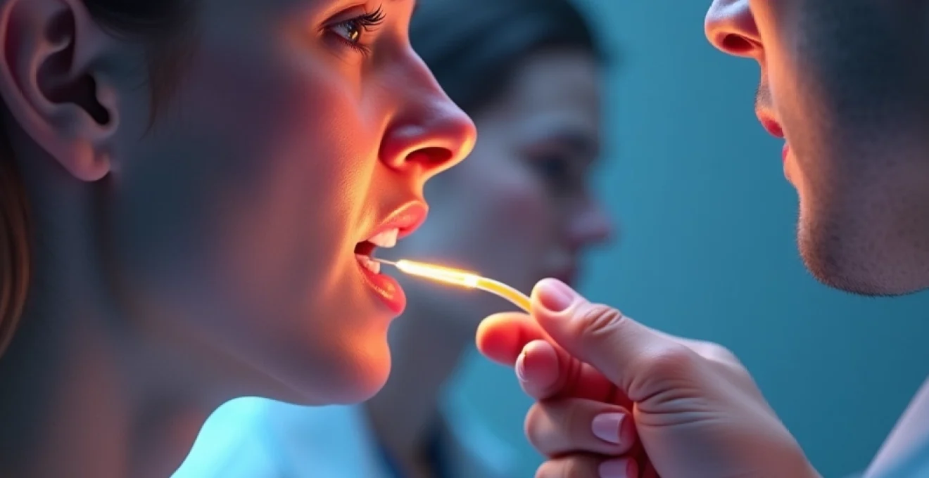
Local anaesthetic injections, particularly for dental procedures, can rarely cause temporary facial nerve paralysis through direct injection into the parotid gland or adjacent neural structures. This iatrogenic complication typically resolves spontaneously as the anaesthetic effect wears off, but patients may experience several hours of facial asymmetry and speech difficulties. Understanding this potential complication helps both patients and healthcare providers manage expectations and avoid unnecessary investigations for more serious underlying conditions.