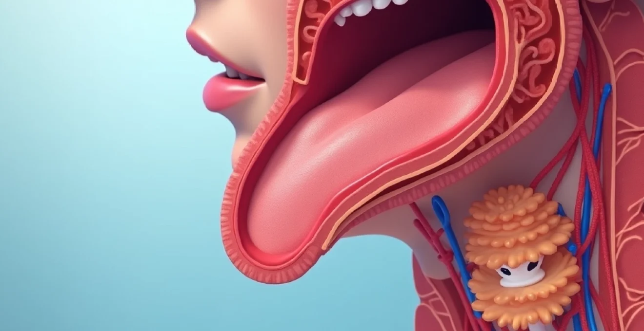
Thyroid colloid cysts represent one of the most common benign thyroid nodules encountered in clinical practice, affecting millions of people worldwide. These fluid-filled structures develop within the thyroid gland when colloid—a protein-rich substance essential for hormone production—accumulates within thyroid follicles. While the vast majority of thyroid colloid cysts remain harmless and asymptomatic throughout a person’s lifetime, understanding their formation mechanisms, diagnostic characteristics, and management options proves crucial for both patients and healthcare providers navigating thyroid health concerns.
Thyroid colloid cyst pathophysiology and formation mechanisms
The development of thyroid colloid cysts involves complex cellular processes that begin at the microscopic level within thyroid follicles. These spherical structures serve as the basic functional units of the thyroid gland, where thyroid hormones are synthesised and stored. Under normal circumstances, follicular cells produce thyroglobulin, which combines with iodine to form the precursors of thyroid hormones T3 and T4.
Follicular epithelium dysfunction and colloid accumulation processes
Follicular epithelium dysfunction represents the primary initiating factor in colloid cyst formation. When the delicate balance between colloid production and hormone synthesis becomes disrupted, excessive colloid begins accumulating within individual follicles. This accumulation occurs gradually, often over months or years, as the follicular cells continue producing thyroglobulin at normal or increased rates while hormone extraction decreases significantly.
The epithelial cells lining the follicles may undergo degenerative changes due to various factors including oxidative stress, inflammatory processes, or genetic predisposition. These cellular changes impair the normal reabsorption mechanisms that would typically prevent excessive colloid build-up, creating an environment conducive to cyst formation.
Thyroglobulin protein aggregation within cystic structures
Thyroglobulin protein aggregation plays a pivotal role in transforming simple colloid accumulation into clinically significant cystic lesions. As colloid density increases within affected follicles, protein molecules begin forming complex aggregates that resist normal enzymatic breakdown processes. These aggregated proteins create a thick, viscous substance that distinguishes colloid cysts from other fluid-filled thyroid lesions.
The protein aggregation process also influences the cyst’s imaging characteristics, particularly on ultrasound examinations where colloid cysts typically demonstrate a characteristic “comet-tail” artifact. This unique appearance results from the acoustic properties of aggregated thyroglobulin proteins, which scatter ultrasound waves in distinctive patterns that experienced radiologists can readily identify.
Iodine deficiency impact on colloid cyst development
Iodine deficiency significantly influences thyroid colloid cyst development through multiple interconnected pathways. When dietary iodine intake falls below optimal levels, the thyroid gland compensates by increasing thyroglobulin production in an attempt to capture available iodine more efficiently. This compensatory mechanism often leads to excessive colloid accumulation within follicles, particularly in geographic regions where iodine deficiency remains endemic.
The relationship between iodine status and cyst formation becomes particularly evident in populations transitioning from iodine-deficient to iodine-sufficient diets. During this transition period, previously enlarged follicles filled with poorly iodinated colloid may undergo cystic degeneration as normal hormone synthesis patterns gradually restore.
Nodular goitre progression to cystic degeneration
Nodular goitre progression represents another significant pathway through which thyroid colloid cysts develop. Long-standing multinodular goitres frequently contain areas of follicular hyperplasia that eventually undergo degenerative changes. These degenerative processes include follicular cell death, hemorrhage, and subsequent colloid cyst formation within the pre-existing nodular tissue.
The progression from solid nodular tissue to cystic degeneration typically occurs over extended periods, often decades. During this transformation, areas of the nodule may become increasingly fluid-filled while maintaining solid components around the periphery. This mixed solid-cystic appearance poses unique diagnostic challenges, as the solid components require careful evaluation to exclude malignancy.
Clinical presentation and diagnostic imaging characteristics
Most patients with thyroid colloid cysts remain completely asymptomatic, with these lesions discovered incidentally during routine physical examinations or imaging studies performed for unrelated conditions. However, larger cysts exceeding 3-4 centimetres in diameter may produce noticeable symptoms including neck fullness, visible swelling, or mild discomfort during swallowing. The clinical presentation varies significantly depending on the cyst’s size, location within the thyroid gland, and proximity to surrounding structures.
Understanding the diverse imaging characteristics of thyroid colloid cysts proves essential for accurate diagnosis and appropriate patient management, as these features help distinguish benign cysts from potentially malignant lesions.
Ultrasonographic features using High-Resolution doppler assessment
High-resolution ultrasonography serves as the primary diagnostic tool for evaluating thyroid colloid cysts, offering detailed visualisation of internal structure and vascular characteristics. Typical colloid cysts demonstrate predominantly anechoic or hypoechoic internal architecture with well-defined margins and posterior acoustic enhancement. The characteristic “comet-tail” artifact appears as bright, linear echoes extending posteriorly from small reflective foci within the cyst.
Doppler assessment reveals minimal or absent internal vascularity within pure colloid cysts, distinguishing them from hypervascular solid nodules that may harbour malignancy. However, peripheral vascularity around the cyst wall remains common and should not raise concern for malignant transformation. Color Doppler evaluation becomes particularly valuable when assessing mixed solid-cystic lesions, where increased vascularity within solid components may warrant further investigation.
Fine needle aspiration cytology findings and bethesda classification
Fine needle aspiration (FNA) cytology of thyroid colloid cysts typically yields abundant colloid material with benign follicular epithelial cells arranged in cohesive clusters. The aspirated fluid appears thick and viscous, often requiring multiple attempts to obtain adequate cellular material for diagnostic evaluation. Cytological examination reveals large sheets of colloid with embedded macrophages and occasional follicular cells showing reactive changes.
According to the Bethesda System for Reporting Thyroid Cytopathology, pure colloid cysts generally fall into Category II (benign) when adequate cellular material is obtained. However, cysts with mixed solid components may require classification as Category III (atypia of undetermined significance) or higher categories if concerning cellular features are present. The cytological findings must be interpreted within the clinical context and imaging characteristics to ensure appropriate patient management.
Computed tomography hounsfield unit measurements
Computed tomography (CT) evaluation of thyroid colloid cysts reveals characteristic density measurements that aid in diagnostic confirmation. Pure colloid cysts typically demonstrate Hounsfield unit values ranging from 20 to 80 HU, reflecting the protein-rich nature of the colloid contents. These measurements fall between simple fluid collections (near 0 HU) and solid tissue masses (typically above 100 HU).
The higher attenuation values compared to simple cysts result from the concentrated thyroglobulin proteins within the colloid material. CT imaging becomes particularly valuable when evaluating large cysts that may be causing mass effects on surrounding structures, as it provides excellent anatomical detail of the relationship between the cyst and adjacent critical structures such as the trachea, oesophagus, and great vessels.
Magnetic resonance imaging T1 and T2 weighted signal intensity
Magnetic resonance imaging (MRI) characteristics of thyroid colloid cysts vary depending on the protein concentration and age of the colloid contents. On T1-weighted sequences, colloid cysts may demonstrate variable signal intensity ranging from hypointense to hyperintense compared to surrounding thyroid parenchyma. Fresh colloid typically appears hypointense, while older, more concentrated colloid may show increased T1 signal intensity.
T2-weighted images generally reveal hyperintense signal within colloid cysts, though this may be less pronounced than in simple fluid collections due to the proteinaceous contents. The MRI signal characteristics can help differentiate colloid cysts from other thyroid lesions, particularly when combined with clinical and ultrasonographic findings. MRI proves especially valuable in cases where CT contrast administration is contraindicated or when evaluating cysts in pregnant patients.
Thyroid function tests including TSH and free T4 levels
Laboratory evaluation of patients with thyroid colloid cysts typically reveals normal thyroid function tests, as these benign lesions rarely affect overall hormonal production. Serum thyroid-stimulating hormone (TSH) levels usually remain within the normal reference range of 0.4-4.0 mIU/L, while free thyroxine (free T4) concentrations maintain normal values between 0.8-1.8 ng/dL.
However, large colloid cysts or multiple cysts within the context of multinodular goitre may occasionally cause mild functional abnormalities. Some patients may develop subclinical hypothyroidism with slightly elevated TSH levels if significant functional thyroid tissue is displaced by cystic lesions. Conversely, autonomous functioning nodules that undergo cystic degeneration may initially present with mild hyperthyroidism before returning to euthyroid status as the cyst matures.
Differential diagnosis from malignant thyroid nodules
Distinguishing thyroid colloid cysts from malignant thyroid nodules requires comprehensive evaluation combining clinical assessment, imaging characteristics, and cytological analysis. While pure colloid cysts carry virtually no malignant potential, mixed solid-cystic lesions require careful scrutiny to exclude concurrent malignancy. The differential diagnosis becomes particularly challenging when dealing with complex cystic lesions that contain solid components or demonstrate atypical imaging features.
Accurate differentiation between benign colloid cysts and malignant thyroid lesions depends on systematic evaluation of multiple diagnostic parameters, ensuring appropriate treatment decisions and patient outcomes.
Papillary thyroid carcinoma cystic variant distinction
Papillary thyroid carcinoma can occasionally present with significant cystic components, creating diagnostic challenges when attempting to distinguish it from benign colloid cysts. Cystic papillary carcinomas typically demonstrate thick, irregular walls with internal solid components showing increased vascularity on Doppler examination. The solid portions often contain characteristic microcalcifications that appear as punctate echogenic foci without posterior acoustic shadowing.
Cytological evaluation of cystic papillary carcinomas reveals malignant epithelial cells with nuclear features including enlargement, overlapping, grooves, and pseudoinclusions. These cellular characteristics contrast sharply with the benign follicular cells found in colloid cysts. Immunocytochemical staining may be employed in challenging cases to identify specific markers associated with papillary carcinoma, such as cytokeratin 19 and HBME-1.
Follicular neoplasm exclusion through cytological analysis
Follicular neoplasms, including follicular adenomas and follicular carcinomas, may undergo cystic degeneration, necessitating careful differentiation from primary colloid cysts. The cytological appearance of degenerating follicular neoplasms typically includes increased cellularity with follicular epithelial cells showing architectural abnormalities and nuclear atypia. These features contrast with the sparse, benign-appearing cells found in true colloid cysts.
Follicular neoplasms often maintain areas of solid tissue even when extensive cystic degeneration occurs. Ultrasonographic examination reveals these solid components as hypoechoic areas with irregular borders and potential vascularity. The presence of a thick, irregular capsule around cystic areas may suggest follicular neoplasm rather than simple colloid cyst formation.
Anaplastic carcinoma rapid growth pattern comparison
Anaplastic thyroid carcinoma represents the most aggressive form of thyroid cancer and can occasionally develop cystic areas due to central necrosis and tissue breakdown. However, the clinical presentation differs dramatically from benign colloid cysts. Anaplastic carcinomas demonstrate rapid growth over weeks to months, often accompanied by compressive symptoms, voice changes, and palpable lymphadenopathy.
The imaging characteristics of anaplastic carcinoma with cystic components show marked contrast enhancement of solid portions, irregular margins, and evidence of local invasion into surrounding structures. The clinical timeline provides crucial diagnostic information , as colloid cysts typically develop slowly over years, while anaplastic carcinomas progress rapidly with devastating clinical consequences.
Medullary thyroid cancer calcitonin level assessment
Medullary thyroid carcinoma (MTC) occasionally presents with cystic degeneration, particularly in larger tumors or those arising from hereditary syndromes. Serum calcitonin measurement serves as a crucial diagnostic tool for identifying MTC, as levels typically exceed 100 pg/mL in patients with clinically significant disease. Normal calcitonin levels (less than 10 pg/mL in men and less than 5 pg/mL in women) effectively exclude MTC as a diagnostic consideration.
Additional biochemical markers including carcinoembryonic antigen (CEA) may be elevated in MTC patients, while genetic testing for RET proto-oncogene mutations becomes appropriate in cases with confirmed or suspected hereditary forms. The combination of biochemical markers and genetic analysis provides definitive differentiation between MTC and benign colloid cysts.
Conservative management protocols and monitoring strategies
Conservative management represents the standard approach for asymptomatic thyroid colloid cysts, particularly those smaller than 4 centimeters in diameter with benign cytological characteristics. This strategy involves regular clinical follow-up combined with serial ultrasonographic monitoring to detect any significant changes in cyst size or internal architecture. The monitoring protocol typically includes initial follow-up examination at 6-12 months, followed by annual assessments if the cyst remains stable.
Patient education plays a vital role in successful conservative management, as individuals need to understand the benign nature of their condition while recognising symptoms that warrant immediate medical attention. Patients should be advised to report rapid cyst enlargement, development of compressive symptoms such as difficulty swallowing or breathing, or changes in voice quality. Regular thyroid function monitoring ensures early detection of any functional abnormalities that might develop over time.
Documentation of cyst characteristics through standardised ultrasound reporting facilitates accurate comparison during follow-up examinations. This includes measurements of cyst dimensions, assessment of wall thickness, evaluation of internal contents, and documentation of any solid components. Photographic documentation of ultrasound images provides valuable reference points for detecting subtle changes during subsequent examinations.
Some patients may benefit from therapeutic aspiration when cysts cause cosmetic concerns or mild compressive symptoms. However, aspiration procedures carry a high recurrence rate, with most cysts refilling within several months. The aspirated fluid should be submitted for cytological examination to confirm the benign nature of the lesion and exclude malignant cells that might indicate concurrent malignancy.
Surgical intervention indications and thyroidectomy techniques
Surgical intervention for thyroid colloid cysts becomes necessary when conservative management fails to address patient symptoms or when diagnostic uncertainty persists regarding the lesion’s nature. Primary indications for surgery include compressive symptoms affecting breathing or swallowing, cosmetic concerns in visible cysts, recurrent cyst formation after multiple aspirations, or the presence of concerning solid components that cannot be adequately evaluated through cytological sampling.
The extent of surgical intervention depends on multiple factors including cyst size, location within the thyroid gland, bilateral disease presence, and patient age and comorbidities. Hemithyroidectomy (lobectomy) represents the preferred approach for unilateral cysts confined to one thyroid lobe, as this technique preserves contralateral thyroid function while achieving complete cyst removal. Total thyroidectomy may be considered for bilateral disease or when malignancy cannot be definitively excluded preoperatively.
Modern surgical techniques emphasise nerve preservation and minimally invasive approaches when anatomically feasible. Intraoperative nerve monitoring helps identify and protect the recurrent laryngeal nerves, reducing the risk of postoperative voice changes. Video-
assisted ultrasound guidance ensures precise nodule localisation during thyroidectomy procedures, particularly beneficial when managing smaller cysts that may be difficult to palpate intraoperatively.
Postoperative care protocols focus on monitoring for potential complications including bleeding, infection, and transient hypoparathyroidism. Most patients can expect to return home within 24-48 hours following uncomplicated thyroid surgery, with activity restrictions limited to avoiding heavy lifting for approximately two weeks. Regular follow-up appointments allow surgeons to assess wound healing, monitor calcium levels, and evaluate voice function to ensure optimal recovery outcomes.
The decision regarding thyroid hormone replacement therapy depends on the extent of surgical resection and remaining functional thyroid tissue. Patients undergoing hemithyroidectomy for benign colloid cysts often maintain normal thyroid function postoperatively, though some may develop subclinical hypothyroidism requiring levothyroxine supplementation. Those requiring total thyroidectomy will need lifelong thyroid hormone replacement with careful dose titration based on serial TSH measurements.
Long-term prognosis and recurrence risk assessment
The long-term prognosis for patients with thyroid colloid cysts remains excellent, with the vast majority experiencing no adverse health consequences throughout their lifetime. Pure colloid cysts carry essentially zero malignant transformation risk, providing substantial reassurance for patients concerned about cancer development. Studies tracking patients over decades consistently demonstrate that properly diagnosed colloid cysts maintain their benign characteristics without progression to malignancy.
Recurrence rates following complete surgical excision approach zero, as the removal of affected thyroid tissue eliminates the anatomical substrate necessary for cyst reformation. However, patients may develop new colloid cysts in remaining thyroid tissue, particularly those with predisposing factors such as iodine deficiency or genetic susceptibility to thyroid nodule formation. These new lesions represent de novo cyst development rather than true recurrence of the original lesion.
Long-term monitoring strategies vary based on the management approach selected and individual patient factors. Those managed conservatively require ongoing surveillance through periodic clinical examinations and ultrasonographic assessments, typically at 12-24 month intervals once cyst stability has been established. Patients who undergo surgical treatment generally require less intensive monitoring, with annual thyroid function testing sufficient for most individuals, particularly those maintaining normal TSH levels consistently.
Quality of life considerations play an important role in long-term outcomes assessment, as many patients experience significant anxiety relief following definitive diagnosis and appropriate management of their thyroid cysts. Educational interventions addressing the benign nature of colloid cysts help reduce unnecessary healthcare utilisation and improve patient satisfaction with their chosen management strategy.
Factors influencing long-term prognosis include patient age at diagnosis, cyst size and location, presence of concurrent thyroid conditions, and adherence to recommended follow-up protocols. Younger patients typically demonstrate more stable disease courses, while elderly individuals may experience gradual cyst enlargement due to age-related changes in thyroid architecture. The presence of multinodular goitre may increase the likelihood of developing additional thyroid nodules over time, though this does not adversely affect the prognosis of existing colloid cysts.
Risk stratification models help clinicians identify patients requiring more intensive monitoring or earlier intervention. High-risk features include rapid cyst growth, development of solid components, changes in ultrasonographic characteristics suggesting malignant transformation, or emergence of compressive symptoms. Conversely, stable cysts maintaining consistent imaging characteristics over extended periods can be managed with progressively longer monitoring intervals, reducing both healthcare costs and patient burden while maintaining appropriate safety margins.